Approaching abdominal pain in a female patient: What's the best diagnostic test?
Approaching abdominal pain in a female patient: What's the best diagnostic test?
Authors: David Ledric MD, MEd, Assistant Director of Emergency Medicine Residency, St. Vincent Mercy Medical Center, Toledo, Ohio; Amanda Klukowski, DO, Emergency Medicine Resident, St. Vincent Mercy Medical Center, Toledo, Ohio
Peer Reviewer: Grant S. Lipman, MD, Clinical Instructor of Surgery, Division of Emergency Medicine, Stanford University School of Medicine, Palo Alto, California
Introduction
Abdominal pain is a common chief complaint in the emergency department (ED), and it is a notoriously difficult one to accurately diagnose. While giving a specific reason for the patient's pain is not always possible, the ED physician has to be able to accurately evaluate the patient for a possible surgical problem. In women this becomes difficult in that it can take hours to decide whether it is an obstetric, gynecologic, or gastrointestinal (GI) medical or surgical issue. Once the patient's pregnancy status is determined, the differential list is still daunting. When the presentation of abdominal pain is atypical, the ED physician is faced with choosing the best diagnostic tool to evaluate the pain symptoms among a myriad of laboratory and imaging studies.
Conventional wisdom suggests that women with suspected gynecologic issues should receive an ultrasound study first. Does this still hold? Atypical presentations of appendicitis are best diagnosed with computed tomography (CT). But the questions remain: What type of CT to order, which imaging study best differentiates GI from gynecologic issues, how does this affect the pregnant patient, what is the role of magnetic resonance imaging (MRI), and is it safe to discharge a patient with ongoing pain?
This paper will focus on imaging controversies and challenges in selecting a diagnostic modality. Articles in this summary review the most commonly used imaging studies available in the ED for evaluating abdominal pain. In addition, articles were reviewed that expand upon the determining factors of these modalities, including accuracy, radiation dose, cost, time, and effect on length of stay. Most of the literature reflects studies or essays from the last 2 years, with exception of 1 clinically relevant article that examines the risk to the fetus of ionizing radiation. The objective of this summary is to provide the reader with the most up-to-date information regarding diagnostic studies in women with atypical abdominal pain and aid the provider with an answer to the question: What imaging study is indicated in women with abdominal pain?
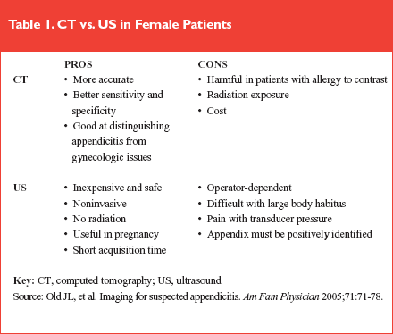
Advantages and disadvantages of CT and US
Source: Old JL, et al. Imaging for suspected appendicitis. Am Fam Physician 2005;71:71-78.
It may be argued that the surgical consultant should be immediately notified when a patient presents with a high clinical suspicion of acute appendicitis, and that imaging studies will only delay the diagnosis. This review article identifies through an evidence-based approach that right lower quadrant pain, anorexia, nausea, low-grade fever, and vomiting are the top 5 criteria warranting a surgical consult. Abdominal x-rays are frequently inconclusive and should be avoided in women with abdominal pain.
Ultrasound (US) is discussed as a safe, noninvasive means of visualizing the pelvic organs and appendix, but has significant limitations for excluding the diagnosis of appendicitis. (See Table 1.) CT has greater sensitivity and specificity for the diagnosis of appendicitis and may even be able to suggest the diagnosis by finding periappendiceal inflammatory changes. In contrast, US can only exclude the diagnosis of appendicitis if a normal appendix is visualized. This is dependent upon the patient's body habitus, appendix location, and the operator's skill.
 |
The article delineates 3 types of CT — the unenhanced CT of the abdomen and pelvis, an oral or intravenous (IV) contrast-enhanced CT of the abdomen and pelvis, and the focused appendiceal CT using rectally administered contrast and a limited scan. This final option is discouraged, as it gives a narrow field of view and will miss alternative abdominal pathology. The conclusion is that the CT scan is the best tool to differentiate bowel from gynecologic etiologies of disease, but a transvaginal US is indicated to further evaluate gynecologic etiologies once a GI etiology has been eliminated.
Commentary
The authors provide a concise summary regarding the difficulty in definitively diagnosing appendicitis. Women not only have a much broader differential diagnosis but also a higher rate of appendicitis and a higher likelihood of an atypical presentation. They cite the surgical and epidemiological literature to point out that nearly 260,000 appendectomies are performed yearly and that women have a higher rate of negative laparotomies than men. Approximately 15% of removed appendixes showed no disease, but in women younger than 40 years of age, as many as 45% in 1 series were normal.
In obvious cases, immediate surgical referral is appropriate, but most cases are atypical and an ED physician must be able to understand the advantages and disadvantages of various imaging studies. This may be dependent on any number of factors, which include the patient, the presentation, the consultant, and the institutional availability of resources. Although this article is primarily a review, the literature it cites and conclusions it makes are very appropriate and practical for any ED physician. This article sets a solid foundation for the studies that follow.
The necessity of contrast
Source: in't Hof K, et al. Surgical validation of unenhanced helical computed tomography in acute appendicitis. Br J Surg 2004;91:1641-1645.
This European prospective study attempts to answer if an unenhanced helical CT is accurate in diagnosing appendicitis, as well as identifying other sources of pathology. In this study, 103 patients with right lower quadrant pain were diagnosed with an acute abdomen by senior surgeons who made the decision to take the patient to the operating room with the diagnosis of an acute abdomen based on clinical suspicion prior to any CT scanning. While waiting for the operating room, all these patients underwent a non-contrast imaging study, and this result was recorded and matched to the patient's operative findings.
The results found that 4 more patients were diagnosed by laparoscopy than by CT (87 vs 83) giving the CT scan a sensitivity of 84.5%. Twenty patients did not demonstrate signs of appendicitis on CT, including the 4 that had appendicitis diagnosed by laparoscopy. The CT scan diagnosed alternative abdominal pathology in 12 patients, which was confirmed on laparoscopy. Finally, at a 6-week follow-up of the patients, no additional pathology was identified.
The authors state that had they changed the intended treatment based on the CT findings, a McBurney incision in 9 women and 5 men would have been prevented. They go on to further point out that even if the decision to go to the operating room wasn't changed, the surgical approach would have. They concluded that a plain helical CT in patients with suspected acute appendicitis provides an accurate diagnosis without the disadvantages of contrast enhancement.
Commentary
Controversy still exists as to what type of CT to order in patients with suspected appendicitis. This article suggests that an unenhanced CT of the abdomen and pelvis is acceptable and points to the advantages of time, comfort and cost over enhanced CT. Interestingly, in this series for the diagnosis of appendicitis, use of IV and rectal contrast accounts for 25% of the total costs of CT. The advantages of not having to administer contrast are clear: decreased amount of time for administration of IV, oral, or rectal contrast improved flow of the ED and potentially fewer patient complications secondary to an earlier diagnosis. Furthermore, unenhanced CT is a good alternative for patients with an allergy to contrast.
There are a number of confounding factors with this study. The conclusion reached may be overstated. In this study, the CT sensitivity for appendicitis was only 85% while most other literature puts the sensitivity into the 90% or better range. It is not known if the 4 missed cases would have been discovered had contrast been given. Also, all the enrolled patients were considered to have a surgical diagnosis. The included patients may be different than the patients with a less typical presentation; in other words, those patients for whom we are most dependent on imaging studies. For the purposes of this review, another problem is that only 39 female patients were included. While CT was able to identify other sources of right lower quadrant pain in 12 out of 20 patients, it is not clear how often this included a gynecologic etiology. The value of contrast in differentiating gynecologic and GI pathology is not clear from this paper alone.
Avoiding false negative CT scans
Source: Levine CD, et al. Why we miss the diagnosis of appendicitis on abdominal CT: Evaluation of imaging features of appendicitis incorrectly diagnosed on CT. Am J Roentgenol 2005;184:P855-859.
Patients with a negative or normal CT may have appendicitis. In an attempt to determine the factors that resulted in CT study missing the diagnosis, Dr. Levine and colleagues retrospectively studied 24 patients with surgically proven appendicitis that had been diagnosed inappropriately as having a negative abdominal CT scan (designated the "case group"). They compared this to a control group of 36 patients diagnosed with appendicitis on both the surgical and preoperative CT reports from the same time period. Images and patient information were reviewed independently by 2 experienced, blinded radiologists.
The review showed that some differences exist between the groups. Patients with a missed diagnosis of appendicitis tended to be leaner, having significantly less intraperitoneal fat (only 1 patient in the case group had significant intraperitoneal fat compared to 17 in the control group). In the cases of missed appendicitis, the radiologist was much less likely to have a clinical history of right lower quadrant abdominal pain or rule-out appendicitis (33% case group vs. 89% in control). Radiologic factors that were correlated with an accurate diagnosis included a visualized appendix, cecal wall thickening, pericecal fat, appendiceal inflammation, lack of intraperitoneal free fluid, lack of perforation, and adequate distal small bowel opacification.
Commentary
This study specifically reviewed cases with a missed diagnosis, a unique way of enrolling patients. While it is obviously written by and intended for radiologists, there are valuable lessons for the ED physician. Unlike the surgeons in the prior paper, the authors felt that oral contrast does improve the ability to make a diagnosis. It is harder to make the diagnosis in women and anything we do to improve the accuracy of the diagnostic testing and help our communication with consultants will help our treatment. They also suggest that in slim patients the diagnosis is more difficult due to the lack of the natural contrast provided by intraperitoneal fat. Most important, providing a clinical history to the radiologist significantly improves the ability to diagnose appendicitis by CT.
Will CT change disposition?
Source: Nagurney JT, et al. Use of diagnostic testing in the emergency department for patients presenting with non-traumatic abdominal pain. J Emerg Med 2003;25:363-371.
This prospective study of 124 patients (76 women) examined laboratory and imaging studies and their value as an adjunct for the diagnosis of non-traumatic abdominal or flank pain in an urban university hospital that sees 73,000 patients annually. Both residents and attendings were asked to group patients into 1 of 6 disposition groups prior to any diagnostic studies, then they were asked which study was most useful for establishing a final diagnosis and disposition. The groups included: home with specific therapy, home with nonspecific therapy, admit for immediate surgery, admit for possible surgery, admit for procedure, and admit for observation. All patients received routine blood tests and 76% received urine tests. The attendings oversaw all further additions of diagnostic tests based on the initial impression following the provided history and physical. Thirty-nine percent of the patients received a CT of the abdomen and pelvis, and 25% received US only. Additional tests were ordered as deemed necessary. CT accounted for 40% of additional testing, and US accounted for 10%. The most frequent indications for further testing included unexpected elevation of the white blood cell count and normal results on initial testing. A change in disposition occurred 43% of the time with CT, versus only 10% with US. Follow-up was achieved by reviewing inpatient charts and calling outpatients by telephone. As a result, the authors indicate that urinalysis and CT were the most useful tests for identifying the cause of non-traumatic abdominal pain.
Commentary
This paper looked more at physician behavior than the accuracy of the CT scan or US. It does give insight into the current practice of emergency medicine in a teaching institution and a possible reflection of community practice, as well. The study is limited by the population size and that the patients were enrolled in a convenience fashion; however, this does not automatically negate the results. The authors readily admit that this was a pilot study but point out its value in looking at ED throughput times and how testing can change the length of patient stays.
It is not surprising that the CT scan was most often responsible for changing the disposition. It also shows the limitations in the value of ultrasonography. One may argue that a test that changes your disposition more than 40% of the time certainly has value. Unfortunately, the study focuses on the results and performance of the imaging study, but has yet to find any objective criteria on when to order it.
How should we proceed?
Source: Riddell AM, Khalili K. Assessment of acute abdominal pain: Utility of a second cross-sectional imaging examination. Radiology 2006;238:571-577.
Sometimes in clinical practice, the emergency medicine physician decides to follow an initial CT with a US (or an initial US with a CT), and sometimes a radiologist recommends a follow-up imaging protocol. This paper examined the utilization of a second imaging study in providing additional information or altering the management of the patient. This retrospective study enrolled 149 adult patients (52% women) who had undergone both CT and US, then had follow-up with a definitive diagnosis. Of these patients, the second imaging study agreed with the first and provided additional information in one-third of the patients. In two-thirds of the patients, the second study agreed with the first but provided no additional information. In only 6.7% of the patients the second study contradicted the first. The second imaging study led to a change in treatment in only 9% of the patients.
The primary reasons for following the US with a CT study were a failed attempt to visualize the appendix or identification of hydronephrosis without a specific cause. The primary reasons for following a CT scan with a US were to further evaluate cholecystitis or renal lesions. The primary finding of this paper was that the yield of the second imaging study was much higher when recommended by the radiologist than it was when ordered independently by the primary physician.
Commentary
In most institutions, the ED physician takes responsibility for interpreting the studies (e.g., electrocardiograms, x-rays, etc.) that are ordered. The exception to this is with abdominal imaging in which the radiology and emergency departments must collaborate. The radiologist authors reach the same conclusion as Dr. Levine and colleagues, that the diagnostic accuracy of the study is significantly improved when the emergency medicine physician and radiologist are able to communicate and share clinical information. In their discussion, the authors state, "the interaction and consultation between the emergency and imaging departments is of utmost importance and should be simplified."
The authors felt that the radiologist was able to make the best suggestion as to when a second study should be ordered but stopped short of claiming a "gatekeeper" role. Acknowledging that useful information was sometimes obtained when the ED was ordering the studies independently, this article highlights the need for emergency medicine physicians to have a good working relationship with their radiologists and to involve them appropriately when they are ordering follow-up studies.
Reducing the negative appendectomy rate
Source: Rosengren D, et al. Radiological imaging to improve the emergency department diagnosis of acute appendicitis. Emerg Med Australas 2004;16:410-416.
This study reviewed the charts of patients who underwent appendectomy during a 12-month period, to determine the utilization and accuracy of US, CT, and abdominal x-ray and the impact of imaging studies on negative appendectomy rate, time, and cost. This Australian study was a retrospective review of 240 patients in a metropolitan teaching hospital.
The likelihood ratio for appendicitis in patients (both sexes, age 14–78) with a normal US was 0.83 (CI 0.56–1.24) and for a normal CT was 0.08 (CI 0.01–0.60). The authors concluded, "With the increasing utilization of CT, a reduction in the negative appendectomy rate by 40–70% to a figure of 4–7 % could be achievable." The paper identified the impact of diagnostic studies on delays to surgical intervention as 9.5 hours with CT and 6 with US.
Commentary
This paper brings a different perspective in that it occurred at an institution that was very comfortable with US and less so with CT scanning. US is heavily utilized in Australia compared to Europe and the United States, and the authors wondered about the effect of the imaging study on the accuracy of their diagnosis.
Radiologists performed more than 4 times the number of US studies as CT scans. In fact, given the small number of CT scans, the author's conclusion seems over-enthusiastic. Other studies demonstrate a negative appendectomy rate of around 8% in patients evaluated by CT scan. Dr. Rosengren and colleagues see a patient population that has a 22% negative appendectomy rate in females with atypical presentations. Given this high rate of negative appendectomies, incorporating CT scanning may likely make a significant difference.
In answer to the question, it appears CT scanning will reduce the negative appendectomy rate but we cannot expect it to disappear. Patients with an equivocal CT scan (i.e., appendix not clearly visualized) will still have a reasonable chance of having appendicitis. CT scanning will have its greatest effect in atypical presentations.
CT diagnosis of gynecologic issues
Source: Bennett GL, et al. CT of the acute abdomen: Gynecologic etiologies. Abdom Imaging 2003;28:416-432.
This pictorial essay begins by suggesting that the previously held belief that US should be used as the first-line study in a patient with abdominal pain of a suspected gynecologic etiology is wrong. As long as the patient is not pregnant, this article argues that a CT is more comprehensive and adept at distinguishing between the bowel and adnexa with the addition of oral contrast as well as discerning the uterus from the adnexa with IV contrast: "CT offers a more comprehensive evaluation than US because of the larger scan field of view." This article demonstrates that CT can be used as a first-line in patients in whom US is difficult due to body habitus, or as a complement to US. When used as a complement, CT helps with identifying inflammatory disease and with staging metastatic disease or the spread of endometriosis.
Commentary
This article appears in the radiologic literature and highlights the continual advances that are occurring in subspecialties. Although this article is an essay and not a scientific study, it demonstrates the rapidly changing diagnostic accuracy of CT as an imaging modality. Awareness of these advances will significantly benefit the ED physician.
Ultrasound is still predominantly suggested as first-line in clear-cut cases of gynecologic etiologies, particularly ovarian torsion. In many cases, however, the etiology is unclear and the CT scan may have a better chance of correctly identifying the pathology. The CT gives one a more comprehensive view of the region under study, is able to better distinguish between the bowel and adnexa, and, especially if used with an appropriate clinical history, is less likely to require a follow-up study.
Communication with the radiologist is critical; advances in diagnostic imaging must be carefully correlated with clinical findings and collaboration is critical to facilitate the optimal diagnostic test.
What is the risk of ionizing radiation to the fetus?
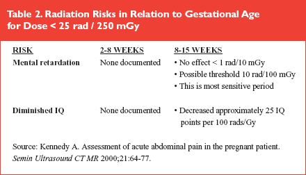
Source: Kennedy A. Assessment of acute abdominal pain in the pregnant patient. Semin Ultrasound CT MR 2000;21:64-77.
This is the oldest of the articles included in this review. It discusses radiation exposure due to the various studies, including the recommendations of the National Council for Radiation Protection (NCRP) and the International Council for Radiation Protection (ICRP).
US has no effect on a fetus and should be the first-line imaging study in the pregnant patient. MRI does not have ionizing radiation but its safety is unproven; it should not be used in the first trimester and only used when US is non-diagnostic or as an alternative to ionizing radiation. Gadolinium crosses the placenta and is contraindicated. When using MRI, US should be used first to determine gestational age.
The NCRP and ICRP guidelines state, "No single diagnostic procedure results in a radiation dose significant enough to threaten the well-being of developing embryo and fetus." They go further to say, "At dose levels below 10 mGy (1 rad) the probability of detectable effect induced by such exposure is so small as to be outweighed by any significant medical benefits." The most sensitive exposure time for embryo and fetus is 8-15 weeks, followed by 16-25 weeks. (See Table 2.)
 |
Regarding CT, Dr. Kennedy asserts that, "In general, the conceptus dose during CT of the abdomen is unlikely to exceed 10 mGy (1 rad) and does not exceed 30 mGy (3 rads) if the fetus is not in view of the beam. When the uterus is directly viewed, the dose will be higher, in the range of 10-40 mGy (1 to 4 rads) and in exceptional circumstances might reach 100 mGy (10 rads)." The approximate radiation dose is 4 rads for abdominopelvic CT, 2.2 rad for pelvic CT, and 50 mrad for an abdominal x-ray. A chest x-ray will give a dose of 8 mrads to the fetus.
Commentary
In pregnant women, not only does the differential diagnosis change but so do the considerations in selecting an appropriate study. It must be remembered that appendicitis is still a significant cause of abdominal pain in pregnancy. While US is the primary imaging modality in pregnancy, it is limited. In some cases, a risk analysis must be done. One must balance the risk of an unnecessary procedure against the potential harm of ionizing radiation in order to make a correct diagnosis.
Indications for MRI in women with abdominal pain
Source: Oto A, et al. Right-lower-quadrant pain and suspected appendicitis in pregnant women: Evaluation with MR imaging—initial experience. Radiology 2005; 234: 445-451.
This study is one of the first to evaluate MRI as a potential imaging modality for pregnant women with suspected appendicitis. The authors point to the advantages of excellent soft tissue resolution and a lack of ionizing radiation. This study was a retrospective review of 23 patients who underwent a non-contrast MRI to evaluate a possible appendicitis. The studies were reviewed by 2 radiologists blinded to the clinical outcome of the patient. All studies were non-contrast studies. Gadolinium crosses the placenta and is contraindicated in pregnancy.
An excellent degree of accuracy was obtained by the radiologists. They were able to identify all the patients with a surgical problem, which included 4 with appendicitis, 3 ovarian torsions, and 2 others with pelvic abscesses not related to appendicitis. They were able to positively identify a normal appendix in 17 of the 19 patients without appendicitis.
Commentary
While it is unlikely that an emergency medicine physician will be ordering an MRI on a pregnant patient in isolation of the radiologic and obstetric consultants, this is an emerging technology that bears watching. In institutions where an MRI is available, the lack of ionizing radiation and ability to have the similar sensitivity and field of view of a CT scan make this a desirable option.
The 2 non-ionizing studies, MRI and US, were not directly compared in this review, but based on previous literature one expects that the MRI would outperform US. Unfortunately, the safety of MRI on the fetus is not fully known. While the authors discourage MRI use in the first trimester, they also cite recent studies as failing to show any experimental or clinical evidence of teratogenic or other adverse effects. Given its limited availability, MRI is unlikely to supplant US or CT, but it is an option that could be considered in limited situations.
Conclusion
It would be difficult and perhaps even inappropriate to come up with hard rules on when to order a CT, US, or MRI on a non-pregnant woman with abdominal pain. The test selection will depend in part on the patient, on the circumstances, and on the institution. From the reviewed articles, however, we may be able to draw some general principles that will guide the clinician in test selection.
In cases of a clear surgical etiology, the appropriate gynecologic, obstetric, or surgical consultant should be immediately notified. Imaging the patient in isolation of the consultant's input will delay the time to the operating room and is more likely to result in a confused diagnosis. As any experienced ED physician knows, there are few cases that are entirely obvious. CT scanning is the test most likely to aid in the disposition. It can distinguish between gynecologic and GI etiologies. It can find unsuspected pathologies and can define the extent of the disease, even in the case of gynecologic problems. There does appear to be a slight advantage in the accuracy of the scan when performed with contrast, and there is definitely an advantage in speaking with the radiologist who will be interpreting the test. CT scans, when properly used, will reduce the number of negative appendectomies. It is important to remember that the scan has excellent — but not perfect — accuracy. In cases where the anatomic structures are not clearly visualized, the scan can only be considered equivocal, and in cases where the clinical suspicion remains high, serial exams may be appropriate.
US does offer advantages in speed and a lack of contrast, but the clinician must be very familiar with the limitations of the diagnostic modality and consider that a nonvisualized appendix is not a negative study. In early pregnancy and in cases of a clear gynecologic etiology, this is the preferred first study. US has only a limited role in the evaluation of appendicitis and in other non-gynecologic etiologies. MRI is another option, depending on institutional availability and the comfort of the radiologists interpreting the images.
Finally, it is important to remember that the treating ED physician must have a good working relationship with the radiologists and surgical consultants. In these cases, the emergency medicine physician may be directing the care early, but will not be the one ultimately responsible for interpreting the images or providing definitive care in the case of surgical pathology.
Abdominal pain is a common chief complaint in the emergency department (ED), and it is a notoriously difficult one to accurately diagnose.Subscribe Now for Access
You have reached your article limit for the month. We hope you found our articles both enjoyable and insightful. For information on new subscriptions, product trials, alternative billing arrangements or group and site discounts please call 800-688-2421. We look forward to having you as a long-term member of the Relias Media community.