Pediatric Circulatory Failure
Authors
Erin Tromble, MD, Resident, Departments of Pediatrics and Emergency Medicine, University of Arizona, Tucson, AZ
Aaron Leetch, MD, Assistant Professor, Departments of Emergency Medicine and Pediatrics; Associate Program Director, Emergency Medicine/Pediatrics Residency, University of Arizona, Tucson, AZ
Dr. Tromble and Dr. Leetch report no financial relationships relevant to this field of study.
Objectives
Etiology
- Know major etiologies of circulatory failure/shock
Pathophysiology
- Understand the pathophysiology of cardiogenic shock
- Understand the pathophysiology of hypovolemic shock
- Understand the pathophysiology of neurogenic shock
- Understand the pathophysiology of distributive shock
Recognition
- Recognize signs and symptoms of compensated shock
- Recognize signs and symptoms of uncompensated shock
Management
- Know the role of crystalloid infusion in the management of shock
- Know the role of pharmacologic therapy for circulatory failure/shock
- Know the role of blood product infusion in the management of shock
Pediatric Cardiogenic Failure/Shock
Etiology
Cardiogenic failure or shock encompasses a wide array of processes that result in insufficient oxygen and nutrient supply to meet the body’s metabolic needs. Shock is defined as insufficient delivery of oxygen or nutrients to tissues. If uncorrected, this state leads to development of anaerobic metabolism,lactate production, metabolic acidosis, and, ultimately, multisystem organ failure and death. Respiratory failure is the most common cause of cardiogenic failure and arrest in children. Other etiologies include sepsis, metabolic derangements, trauma, and intrinsic cardiac disease.
Pathophysiology
Shock ultimately develops with either impairment of cardiac output (CO), systemic vascular resistance (SVR), or both.
There are several categories of shock, which include the following:
- Hypovolemic – Decreased intravascular volume, (either hemorrhagic or non-hemorrhagic). Volume loss results in decreased pre-load and stroke volume, causing decreased cardiac output (CO).
- Septic – Caused by an infectious insult (bacterial, viral, fungal) resulting in inflammatory responses that cause increased metabolic demand and impaired perfusion. Pediatric presentation is variable. The classic adult presentation of low SVR and high CO is present in the minority of pediatric patients.
- Distributive – Results from a peripheral vasodilatory state leading to low SVR. Two major causes include anaphylaxis (allergic reaction leading to massive histamine release) and neurogenic (CNS injury causing loss of autonomic tone).
- Cardiogenic – CO is impaired by myocardial dysfunction, dysrhythmias, or congenital malformations
- Obstructive – Preload is restricted due to obstruction of venous return. Shock develops from decreased CO. Two major causes include tension pneumothorax and pericardial tamponade.
Table 1: Types of Shock
|
Type |
Pathophysiology |
Signs & Symptoms |
|
Hypovolemic |
Intravascular volume loss, ↓ CO |
Tachycardia, ↓ pulses, ↓ capillary refill, hypotension (late in pediatric patients) |
|
Septic |
Infectious insult, varying pediatric presentation of CO and SVR |
Tachycardia, variable blood pressure, ↓ capillary refill |
|
Distributive |
↓ SVR, ↑ CO in anaphylaxis, ↓ CO in neurogenic shock |
Anaphylaxis: angioedema, respiratory distress, tachycardia Neurogenic: normal or low heart rate, hypotension, paralysis |
|
Cardiogenic |
↓ CO, normal or ↑ SVR |
Normal to elevated heart rate, hepatomegaly, cardiomegaly, normal to low blood pressure |
|
Obstructive |
↓ CO, normal or ↑ SVR |
Jugular venous distention, Tension pneumothorax: Absent breath sounds Pericardial Tamponade: Muffled heart sounds |
Pediatric shock is a unique entity, distinct from that of adults, and must be evaluated with attention to the qualities that make pediatric shock.
Table 2: Characteristics of Pediatric Shock
|
CME Question
A 14-year-old boy was the unhelmeted driver of an ATV involved in a collision. The patient was separated 30 feet from the vehicle and found unresponsive on the scene. On arrival to the emergency department, the patient has obvious signs of trauma to the head and face and a heart rate of 166 with a blood pressure of 73/42. What is the most likely explanation for the patient’s vital sign abnormalities?
- Hemorrhagic shock
- Tension pneumothorax
- Loss of autonomic tone
- Adrenal insufficiency
- None of the above
Start or Resume CME Test
(Opens in a new Tab/Window)
Diagnosis
Shock can be challenging to identify in the pediatric patient. Clinical signs are often subtle and may be limited to tachycardia in the early compensated phase.Careful attention to appearance, work of breathing and color may identify early clues. (See Table 3.) Irritability, lethargy, and fluctuations in mental status may indicate poor perfusion. Increased respiratory effort may be due to respiratory distress or due to increased metabolic demand. End-organ perfusion can be assessed in the form of capillary refill (skin), urine output (kidneys), mental status (CNS), and heart rate (cardiac). In the setting of a normal blood pressure, poor perfusion indicates compensated shock, which may quickly progress into decompensated shock without intervention. Hypotension is generally a late, and often ominous, finding.
Table 3: Rapid Assessment of Pediatric Patients (“Pediatric Assessment Triangle”)
|
Appearance |
Level of alertness Interaction Response to stimuli |
|
Work of Breathing |
Respiratory rate Respiratory effort (retractions, nasal flaring, grunting, abdominal breathing) |
|
Color |
Cyanosis Pallor Mottling |
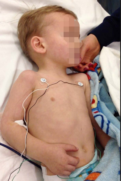
Figure: A Child with Mottling and Poor Perfusion
Diagnostic Evaluation
Laboratory data should be obtained and interpreted in the context of the patient at hand. Arterial or venous blood gas analysis and serum lactate measurement may reveal a metabolic acidosis secondary to hypoxemia and anaerobic metabolism. CBC, comprehensive metabolic profile (CMP), blood glucose level, and coagulation studies may be non-specific but should be interpreted in the context of the clinical picture. Blood and fluid cultures should be obtained, if an infectious etiology is suspected.
Table 4: Classic Laboratory Findings in Shock
|
Classic Laboratory Findings |
|
|
Septic Shock |
WBC: ↑ Lactate: ↑ ScvO2: ↓ Glucose: ↑ |
|
Hypovolemic |
WBC: generally ↑ Lactate: ↑ Hgb/HCT: ↓ (hemorrhagic – may be nl initially) PTT, PT/INR: ↑ (hemorrhagic) Glucose: ↑ |
|
Distributive |
Neurogenic: WBC: generally ↑ Lactate: nl early, ↓ late Anaphylactic: WBC: ↑ Glucose: ↑ |
|
Cardiogenic |
WBC: varies with etiology, but generally ↑ ScvO2: ↓ Lactate: ↑ |
WBC = white blood cell count, ScvO2 = central venous oxygen saturation, Hgb = hemoglobin, HCT = hematocrit, nl = normal, PTT =partial thromboblastin time, PT = prothrombin time, INR = international ratio
Management
Early recognition and aggressive intervention is essential and should be in accordance with the American Heart Association Guidelines for Pediatric Advanced Life Support (PALS). In undifferentiated shock, intervention should include the following:
-
Venous access
- Two proximal large bore IVs.
- Intraosseous access, if the above cannot be established quickly.
- Central venous access may be indicated in advanced, refractory cases of shock.
-
Continuous monitoring
- Patients should be placed on cardiac monitoring throughout the resuscitation to monitor for dysrhythmias, including bradycardia.
- Continuous pulse oximetry can aid in monitoring and titrating respiratory support.
-
Respiratory support:
- Supplemental oxygen should be administered to all patients in shock.
- Consider intubation if there is impaired mental status or concern for airway protection.
- If signs of respiratory failure are present, intubation should not be delayed.
-
Fluid resuscitation
- 20 mL/kg boluses of isotonic fluid, repeated up to 60 mL/kg or more until perfusion improves or hepatomegaly, rales, or other signs of congestive heart failure develop.
- Patients should be frequently reassessed for response to fluids and to guide further resuscitation.
- Blood products should be transfused if there are signs of hemorrhagic shock, symptomatic anemia, or DIC.
-
Vasoactive medications
- Used in the setting of persistent hypotension despite adequate fluid resuscitation.
- Dopamine (starting dose: 5mcg/kg/min) is the recommended first line agent for septic shock. Infusion should be titrated to establish and maintain adequate perfusion.
- Alternative or additive therapy with norepinephrine, epinephrine, milrinone, and vasopressin should be based on the patient’s clinical picture.
-
Adrenal insufficiency
- Hydrocortisone should be administered in catecholamine resistant shock, history of CNS abnormality (hypopituitarism), or history of recent steroid use.
- Hydrocortisone should be given empirically in cases of vasopressor refractory shock.
- Cortisol levels should be checked prior to administration when possible.
Table 5: Pre-ICU Management of Shock Based on Type
|
Septic |
Hypovolemic |
Distributive |
Cardiogenic |
|
|
Endpoints |
• CRT ≤ 2 secs • Normal peripheral and central pulses • Warm extremities • > 1 mL/kg/hr urine output • Normal mental status • Normal mean arterial pressure (MAP) • Normal central venous pressure (CVP) (if available) • Normal lactate level or >10% lactate clearance • Normal glucose • Normal ionized calcium • ScvO2 >70 (if available) |
• CRT ≤ 2 secs • Normal peripheral and central pulses • Warm extremities • > 1 mL/kg/hr urine output • Normal mental status • Normal BP • (Hemorrhagic) Hgb > 7 • (Hemorrhagic) normal coagulation studies |
• CRT ≤ 2secs • Normal peripheral and central pulses • Warm extremities • > 1 mL/kg/hr urine output • Normal mental status • Normal BP |
• CRT ≤ 2secs • Normal peripheral and central pulses • Warm extremities • > 1 mL/kg/hr urine output • Normal mental status • Normal BP
|
|
Monitoring |
• Pulse oximetry • Continuous ECG • BP (NIBP acceptable if pulses palpable) • Temperature • Urine output • Glucose • Ionized calcium • Blood gas |
• Pulse oximetry • Continuous ECG • BP (NIBP acceptable if pulses palpable) • Urine output • Temperature (for development secondary infection or concomitant sepsis) • Hemorrhagic coagulation studies: PT/INR, PTT, fibrinogen, thromboelastography (TEG)
|
• Continuous ECG • BP (NIBP acceptable if pulses palpable) • Urine output • Temperature (for development of secondary infection or concomitant sepsis)
|
• Pulse oximetry • Continuous ECG • BP (NIBP acceptable if pulses palpable) • Urine output • Temperature (for development of secondary infection or concomitant sepsis) |
|
Airway & Breathing |
• Supplemental oxygen • Clinical assessment determines need for mechanical ventilation, but need for 60 mL/kg fluid in first hour is reasonable indication for intubation. 40% of cardiac output is used to power work of breathing – intubation and mechanical ventilation may reverse shock. • Ketamine plus NMJ blocker recommended for intubation. Etomidate is hemodynamically neutral but may cause adrenal insufficiency in septic shock (unclear clinical implications). Other induction agents may cause hypotension. • Fluid resuscitation prior to sedation and positive pressure ventilation may prevent post-intubation hypotension. • Consider concomitant IV inotropes during RSI to prevent cardiovascular collapse. |
• See “Septic” |
• Supplemental oxygen • In anaphylaxis, if signs of significant respiratory distress or airway compromise, intubation should be sought quickly as ability to intubate will rapidly decline with progression of allergic reaction |
• Supplemental oxygen should be used cautiously in patients with congenital cardiac defects as may worsen clinical status • Non-invasive ventilation may be beneficial if patient able to tolerate and alert • If severe shock or altered mental status, mechanical ventilation likely indicated |
|
Circulation |
• IO access if IV access is not obtained within minutes • Fluid bolus (0.9% saline) 20 mL/kg immediately if no hepatomegaly or rales. If hepatomegaly or rales, give fluid bolus cautiously and consider other diagnoses (heart disease) • Repeat fluid bolus to achieve normal CRT and BP. 60 mL/kg or more is often required in pediatric sepsis in the first hour. • If fluid refractory shock start inotrope peripherally (low-dose dopamine or epinephrine) while establishing central access. • Fluid and dopamine refractory shock – add epinephrine (cold shock) or norepinephrine (warm shock) • If shock is vasopressor refractory or if there is clinical suspicion of adrenal insufficiency/crisis, administer hydrocortisone after obtaining random cortisol level. |
• If hemorrhagic, control source of bleeding • Obtain access as described in “septic” • Crystalloid boluses as outlined in “septic” in non-hemorrhagic types. If hemorrhagic, crystalloid and albumin may be used until blood products are available, but blood products should be obtained rapidly. |
• Obtain rapid access and IO if IV cannot be obtained in minutes • Rapid IM epinephrine, followed by IV if needed, in anaphylaxis • Volume may be administered in 20 mL/kg boluses as needed for shock • Vasoactive agents may be necessary • Phenylepherine and norepinephrine are agents of choice in neurogenic shock |
• IO access if IV access is not obtained within minutes • Cautious administration of IV fluid boluses often starting at 10 mL/kg with frequent reassessment for signs of pulmonary edema • Vasoactive agents are generally indicated and include dobutamine, milrinone, and dopamine |
|
Antibiotics |
• Administered within 1 hour • Broad-spectrum empiric antibiotics: Selection will depend on suspected etiology and patient age. (Preterm neonates: Gentamycin and ampicillin; Term Infants: gentamicin or cefotaxime or ceftazidime and ampicillin; Older infants and children:ceftriaxone or an antibiotic with pseudomonal coverage such as pipercillin-tazobactam or cefipime plus vancomycin; Acyclovir should be added if clinical suspicion of HSV) |
• Indicated if concomitant sepsis suspected as described in “Septic” section | • Generally not indicated | • Indicated if infectious source suspected as precipitating factor in acute decompensation and should be administered as described in “Septic” column. |
CME Question
An 8-year-old male with multiple ICU admissions for asthma presents in respiratory distress with tachycardia, hypotension and an infiltrate on chest X-ray. Despite multiple 20mL/kg fluid boluses followed by initiation of dopamine and norepinephrine, he continues to be hypotensive with signs of poor perfusion. What should be the next step in management?
- Initiate vasopressin therapy
- Initiate epinephrine drip
- Administer corticosteroids
- Repeat fluid boluses at 20ml/kg
- Obtain a chest X-ray
Start or Resume CME Test
(Opens in a new Tab/Window)
National Guideline/Academic Resources
http://www.aacn.org/WD/CETests/Media/ACC2344.pdf
http://pediatrics.aappublications.org/content/126/5/e1361.full
References
Dellinger RP, Levy MM, Carlet JM, et al. Surviving Sepsis Campaign: International guidelines for management of severe sepsis and septic shock. Intensive Care Med 2008;34:17-60.
Goldstein B, Zimmermann JJ. New horizons, the science and practice of acute medicine: Critical care of pediatric shock. Crit Care Med 1998;6:S120-S154.
Tabbutt S. Heart failure in pediatric septic shock: Utilizing inotropic support. Crit Care Med2001;29:S231-236.