Spinal Trauma
Module 3: Pediatric Trauma
Authors
Whitney Kiebel, MD, Resident, Departments of Pediatrics and Emergency Medicine, University of Arizona, Tucson, AZ
Aaron Leetch, MD, Assistant Professor, Departments of Emergency Medicine and Pediatrics; Associate Program Director, Emergency Medicine/Pediatrics Residency, University of Arizona, Tucson, AZ
Dr. Kiebel and Dr. Leetch report no financial relationships relevant to this field of study.
Core Content Outline: Cervical Spine Injury
Etiology
-
Understand the most common etiologies of cervical spine injuries in children
-
Know mechanisms and patterns of injury associated with cervical spine injuries in children
Pathophysiology
-
Understand the types and mechanisms of cervical spine and spinal cord injuries
-
Understand age-related differences in cervical spine injuries
-
Understand the mechanism of injury of a Jefferson fracture
-
Differentiate between neurologically stable and unstable cervical spine injuries
Recognition
-
Recognize signs and symptoms of spinal cord injury syndromes (anterior, central, complete, posterior, Brown-Sequard) in children
-
Recognize the signs and symptoms of findings suggestive of cervical spine injury
-
Know indications for radiographic evaluation of cervical and spinal cord injuries Recognize age-based radiologic variants of the spine and be able to differentiate from pathologic cervical spine injuries
-
Know the role of pharmacologic agents in the management of spinal cord injury
Treatment
-
Plan options for stabilization of cervical spine injuries in pediatric patients of different ages
Thoracolumbar Spine Injury
Etiology
-
Know the most common life-threatening causes of thoracolumbar spine injuries in children
Pathophysiology
-
Understand the types and mechanisms of thoracolumbar spine injuries Understand neuroanatomic clinical correlation in thoracolumbar spine injuries
Recognition
-
Know the significance of symptoms and physical examination findings after blunt thoracolumbar trauma
-
Know radiographic evaluation of thoracolumbar spine injuries, and recognize radiologic variants
-
Recognize injuries commonly found in conjunction with thoracolumbar spine injuries
Treatment
-
Plan options for evaluation, stabilization, and management of thoracolumbar spine injuries
Peripheral Nervous System
-
Recognize the signs and symptoms of peripheral nerve injury Plan the management of peripheral nerve and plexus injury
Neck – Blunt Trauma
-
Understand the significance of injuries associated with blunt trauma to the neck Plan the evaluation and management of a child with blunt trauma to the neck
Neck – Penetrating Trauma
-
Plan the evaluation and management of penetrating injury to the neck differentiating by symptoms and location
-
Recognize potential injuries associated with penetrating trauma to the neck
Pediatric Spine Trauma
Etiology
Spinal trauma is relatively uncommon in children. However, spinal injuries should be considered in all children who have sustained head or neck trauma or multiple severe injuries. Upper cervical spine injury (C1-C4) is more common in children < 8 years of age. Adolescents and adults are more likely to suffer lower cervical spine (C5-T1) injuries. Spinal injuries are more common in boys. Motor vehicle accidents in which the victim was unrestrained or improperly restrained are the most common mechanism of spinal injury in children. Falls are the second most common mechanism in patients younger than 8 years old. In older children, sports injuries are the second most common mechanism. Mortality rates for cervical spine injuries ranges from 16-18%. Higher cervical spine injuries, including atlanto-occipital dissociation, are associated with the highest mortality rate.
A higher degree of suspicion for spinal cord injury should be maintained in children who suffer head injury and/or multi-system trauma. Spinal immobilization is essential to prevent morbidity and mortality.
Children with underlying structural abnormalities are more vulnerable to injury and even minor trauma may result in serious injury. Such patients include those with Down’s syndrome (atlanto-occipital instability), a history of previous cervical spine injury or cervical spine arthritis, Klippel-Feil syndrome (congenital fusion of cervical spine), Morquio syndrome (hypoplasia of odontoid), Larsen syndrome (cervical vertebrae hypoplasia, multiple joint dislocations), and any other syndromes associated with spinal abnormalities. In addition, chin trauma, fractures of the posterior teeth, and mandibular condyles may be associated with c-spine injuries.
National Guideline/Academic Resource
http://www.neurosurgery.org/sections/section.aspx?Section=PD&Page=ped_spine.asp
Pathophysiology
There are several differences between the pediatric spine and the adult spine. The age of 8 years traditionally is considered a transition point in anatomic development. Children 8 years of age and younger are more susceptible to upper cervical spine injuries (C1-C3). Older children more commonly suffer injuries in a pattern similar to adults, i.e., C7-T1 region. Reasons for this include:
-
The cervical spine fulcrum is more cephalad (C2-C3) in children < 8 years old. As children age, it progresses more caudally to the C5-C6 region.
-
Children younger than age 8 years have increased laxity of the cervical spine ligaments and musculature, allowing greater mobility in flexion/extension
-
Children younger than age 8 years have increased elasticity of interspinous ligaments, posterior joint capsule, and cartilaginous end plates
-
Wedge-shaped and horizontally oriented vertebral bodies predispose younger patients to injury
-
Intervertebral disc and annulus have high water content and can have longitudinal expansion without rupture during spinal column distraction
-
Children younger than age 8 years have relatively larger and heavier heads.
Four injury patterns are common in children ages 8 years and younger:
-
Fracture with subluxation
-
Fracture without subluxation
-
Subluxation without fracture
-
Spinal cord injury without radiographic abnormality (SCIWORA)
Younger children are susceptible to growth plate and ligamentous injuries. Children have immature growth centers that are susceptible to sheer forces during rapid deceleration or hyperflexion/extension, particularly at the synchondrosis between the odontoid and vertebral body of C2. In young children, a more elastic spinal column can tolerate more distraction before rupture, thus leading to spinal cord injury without obvious bony spinal column injury or ligamentous disruption.
Common Types of Fractures
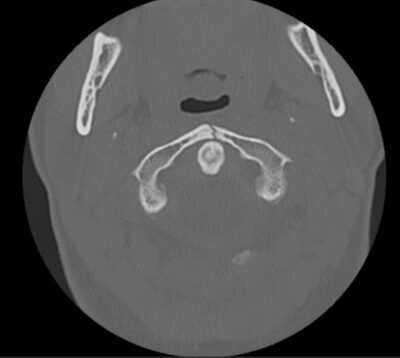
Jefferson fractures refer to a burst fracture of C1 and may involve both the anterior and posterior arches. The mechanism is usually from an axial load (i.e., diving). The fracture is seen on an odontoid view, but this view is difficult to obtain in children < 3 years of age. Typically, there will be a lateral offset of the lateral mass of C1 of > 1 mm from the vertebral body of C2. A pseudo-Jefferson fracture can be seen between 2-6 years of age secondary to the increased growth of the atlas compared to the axis.
Figure: A Patient with a C1 burst fracture

Hangman fracture is a traumatic spondylolithesis of C2, fracture of the pars interarticularis of the axis, horizontal tearing of the C2-C3 disk, and anterior subluxation of C2 on C3. Hangman fractures often result from hyperextension forces. If hyperflexion occurs after hyperextension, C2 may sublux onto C3 and cause spinal cord damage. The subluxation can be mistaken for pseudosubluxation.
Pseudosubluxation is seen in the C2-C3 or C3-C4 region in 25% of children younger than 8 years of age but can persist to age 16 years. On radiographs, evaluate the spinolaminar line, which is drawn from the posterior arches of C1-C4. This should pass within 2 mm of the anterior cortex of the posterior arch of C2 in children with pseudosubluxation. Distances greater than 2mm are usually abnormal.
Atlantoaxial subluxation occurs as a result of movement between C1 and C2 secondary to transverse ligament rupture or a fractured dens. On lateral spine radiographs, there will be a widened predental soft tissue space (normal < 5mm)
Odontoid fractures are the most common cervical spine injuries in children. These fractures may be confused with normal anatomic variations due to synchondrosis between the body and the axis. Type 1 odontoid fractures involve the apex of the dens. Type 2 fractures are through the waist of the dens, and Type 3 extend into the body of C2. Type 2 and 3 are considered unstable and require emergent consultation.
Distraction injuries occur with a longitudinal stress on the cervical column. The most serious type is the occipito-atlanto disociation. On radiographs, there is an increased distance between the occiput and C1 or widening of intervertebral disk space without adjacent compression. These injuries are associated with high cervical spinal cord injuries and are commonly incompatible with long-term survival.
Rotary subluxation involves facet fracture or dislocation. These present with muscle spasm on the sternocleidomastoid on the same side to which the chin points. These injuries are usually associated with a ligamentous injury.
SCIWORA – Spinal Cord Injury Without Radiographic Abnormality – is a clinical-radiological condition that mostly affects children. With recent advances in neuroimaging techniques and increasing availability of MRI, the overall detection rate of SCIWORA has significantly improved. SCIWORA lesions are found mainly in the cervical spine but can also be seen, although much less frequently, in the thoracic or lumbar spine. SCIWORA is responsible for 6 to 19% and 9% to 14% of spinal injuries in children and adults, respectively. The level of spinal cord injury corresponds to the location of these changes.
These injuries can occur through many mechanisms, including hyperextension, hyperflexion, and distraction. In hyperextension injuries, the posterior interlaminar ligaments impinge the spinal cord, whereas, in hyperflexion injuries, the anterior interlaminar ligaments impinge on the cord. Distraction injuries occur when the longitudinal distraction stretches the spinal column beyond the tolerance of the spinal cord. This is seen with rapid acceleration/deceleration forces.
Hematomyelia may occur with these injuries, causing hemorrhage into the spinal cord and severe neurologic injury. Thoracic SCIWORA has been described, but is less common due to the splinting of the ribcage. Patients present with neurologic symptoms and deficits consistent with cord injury but without radiographic abnormalities on imaging (X-ray and CT). Some will only have transient neurologic symptoms and completely recover after 24 hours. Some may not have any neurologic symptoms immediately after the injury, but develop them up to 4 days post-injury.
An MRI is the diagnostic study of choice and a recent systematic review emphasizes the prognostic value of spinal MRI for children with SCIWORA and highlights the role of the MRI classification system in improving the comparability and interpretability.
The prognosis of children with SCIWORA alone is better than that of children with vertebral fractures and neurologic symptoms. Children diagnosed with SCIWORA but with normal MRI finding have substantially more favorable clinical outcomes than those with cervical cord abnormalities on MRI.
National Guideline/Academic Resources
http://www.ncbi.nlm.nih.gov/pmc/articles/PMC3143390/
http://www.ncbi.nlm.nih.gov/pubmed/25807412
http://www.ncbi.nlm.nih.gov/pubmed/24158204
CME Question
11. A 15-year-old presents after suffering an injury while swimming. Bystanders state that he was diving into the pool and may have hit his head on the bottom. On examination, the patient has a normal neurologic examination. He has a large hematoma on the top of his head with a small superficial laceration. He complains of some neck stiffness. What injury are you most concerned for?
-
Hangman’s fracture
-
Jefferson’s Fracture (C1 burst fracture)
-
Subdural hematoma
-
Atlanto-occipital dislocation
-
None of the above
Start or Resume CME Test
(Opens in a new Tab/Window)
CME Question
12. A 7-year-old boy presents to the emergency department after falling from a horse. He suffered a brief loss of consciousness, but woke shortly afterward. He complains of numbness and tingling in his hands, but otherwise has a completely normal neurologic exam. He does not have any spinal tenderness. You obtain a CT of his head and c-spine which were normal. He continues to have numbness and tingling in his hands after several hours in the emergency department. What is the next step in treatment and workup for this patient?
-
Discharge him with a cervical collar and advise him to follow-up with his pediatrician
-
Obtain a repeat CT scan to evaluate for progression of injury
-
Obtain an MRI to evaluate for SCIWORA
-
Arrange for follow-up with a neurosurgeon and discharge home
Start or Resume CME Test
(Opens in a new Tab/Window)
Spinal Cord Syndromes
|
Spinal Cord Syndrome |
Mechanism |
Neurologic deficit |
|
Anterior Cord Syndrome |
Hyperflexion injury and anterior cord compression |
|
|
Central Cord Syndrome |
Hyperextension injuries |
|
|
Brown-Sequard Syndrome |
Hemisection of the cord |
|
|
Horner’s Syndrome |
Injury to cervical paravertebral sympathetic chain and inferior cervical ganglion |
|
|
Cervical Cord Neuropraxia |
Axial loading of neck in flexion or extension (common in football players and increased risk in cervical stenosis) |
|
|
Transection of Spinal Cord |
Severe spinal column disturbance |
|
Diagnosis/Evaluation
Children with spinal cord trauma must be evaluated and recognized promptly. Initial assessment includes the primary and secondary survey with attention to the airway, breathing, and circulation.
-
Hypotension, bradycardia, and temperature instability may be a sign of spinal shock, which results from disruption of the sympathetic chain and unopposed parasympathetic innervation
-
Apnea/hypoventilation may be a result of diaphragmatic paralysis (C3-C5)
Children with fractures typically have pain, muscle spasm, and decreased range of motion. They may complain of transient or persistent paresthesias, paralysis, or weakness. The distribution may vary based on the injury. Some children are unable to express their symptoms, and others may have distracting injuries. Transient burning of the upper extremities may be indicative of a hyperextension injury with central cord contusion.
A thorough neurologic examination should be performed if there is any concern for spinal injury. Spinal immobilization should be maintained and rapid radiologic evaluation should be performed. Nearly 50% of children with cervical cord injuries have neurologic deficits.
|
Neurologic Signs and Symptoms |
|
Abnormal motor exam |
|
|
|
|
|
|
|
Abnormal sensory exam |
|
|
|
|
|
AMS |
|
Neck pain |
|
Torticollis |
|
Limitation of motion |
|
Neck muscle spasm |
|
Neck ecchymosis or swelling |
|
Abnormal or absent reflexes |
|
Transient depression of reflexes below injury |
|
Clonus without rigidity |
|
Diaphragmatic breathing without retractions |
|
Neurogenic shock |
|
Absence of rectal tone |
|
|
Priapism |
|
Absence of bulbocavernosus reflex |
|
|
|
Decreased bladder function |
|
Fecal retention |
|
Unexplained ileus |
|
Autonomic hyperreflexia |
|
BP variability with flushing and sweating |
|
Poikilothermia |
|
Hypothermia or hyperthermia |
Radiographic Imaging
Children with suspected spinal injury should undergo radiographic imaging. Many decision algorithms for cervical spine imaging have been based on adult populations. The NEXUS study included only 2.5% of patients younger than 8 years of age. The Canadian C-Spine Decision Rule was not designed for the evaluation of children. Further challenges in the decision-making process in children include the difficulty in assessing the preverbal, anxious, and upset child.
|
Considerations for cervical spine imaging include: |
|
|
|
|
|
|
In children older than 8 years of age, the NEXUS criteria may be used to determine the need for cervical spine imaging.
|
NEXUS Criteria for Low Probability of Injury |
|
1. No midline tenderness |
|
2. No focal neurologic deficit |
|
3. Normal alertness |
|
4. No intoxication |
|
5. No painful distracting injury |
Radiologic cervical spine evaluation should obtain at least three views of the c-spine (lateral, AP, and, if able, open-mouth odontoid). Cross-table lateral views identify most fractures, dislocations, and subluxations but are not sufficient to exclude injury. AP and odontoid views increase the sensitivity of identifying injuries. CT may be necessary when plain radiographic views cannot be obtained or are indeterminate. Plain radiographs should be utilized first, if possible. Many resources recommend obtaining CT of the upper c-spine in all patients undergoing CT brain/head imaging, particularly those who are unconscious and immobilized. One must weigh the benefits of the study against the increased radiation exposure. Focused imaging may help limit radiation exposure. Neurosurgical guidelines give a Level I recommendation for CT imaging to determine the condyle-C1 interval (CCI) for pediatric patients with potential atlanto-occipital dislocation (AOD).
Interpretation of spinal radiographs in children is difficult and requires an experienced radiologist or provider to interpret the anatomic variations. Some of these include:
-
Cervical lordosis
-
Incomplete ossification of the posterior elements
-
Ligamentous laxity
-
Pseudosubluxation of C2 on C3 or C3 on C4
-
Anterior wedging
-
Disruption of alignment of the four c-spine contour lines (anterior vertebral body line, posterior vertebral body line, spinolaminar line, and tips of the spinous processes)
-
Muscle spasm can disrupt the lordotic curve
-
Pseudosubluxation
-
Ligamentous disruption may alter the line at the tips of spinous processes
-
Unstable occipitoatlantoaxial injury suspected when distance between spinous processes of C1 and C2 is increased
-
-
Predental space: between posterior surface of the anterior arch of C1 and anterior surface of odontoid
-
< 8 years old, 4-5 mm (3 mm in adults)
-
Widening indicates blood or edema
-
Secondary to atlantoaxial instability, Jefferson fracture, or atlantoaxial rotation subluxation
-
-
-
Prevertebral space at C2-C4
-
Should be no more than one-third to one-half the AP diameter of the adjacent vertebral body
-
Widening may indicate hematoma, abscess, or ligamentous injury
-
May appear falsely widened on films obtained during expiration and if the neck is overly flexed
-
Flexion/extension views have limited utility and should not be routinely obtained in the emergency department. They require a cooperative patient able to flex and extend the neck without pain or spasm.
CT scan is an excellent modality for identifying bony abnormalities. MRI is preferred for visualizing soft tissues, ligamentous injuries, spinal cord edema, hemorrhage, compression, and transection.
Management/Treatment
Children with clinical findings suggestive of spinal cord injury should be treated conservatively with immobilization, even in the absence of radiographic abnormalities. Referral to a regional trauma center for pediatric neurosurgical and pediatric orthopedic evaluation in some circumstances is crucial for definitive management. All patients with documented spinal cord injury and neurologic deficit should be admitted to the pediatric intensive care unit.
Steroid therapy is debated, and randomized controlled trials are needed in the pediatric population to assess the advantages of steroid use after SCI in children. The use of steroids in patients is associated with increased infectious risks and no neurological improvements.
National Guideline/Academic Resource
http://www.ncbi.nlm.nih.gov/pubmed/21994079
Some patients may meet criteria for discharge from the emergency department with a padded hard cervical collar. Consider discharge if any of the following are present:
-
Transient symptoms have resolved
-
Normal neurologic examination
-
Normal imaging
-
No other serious injuries
-
Parent or caregiver and patient aware of restriction of any activity that may lead to re-injury
-
Neurosurgical follow-up in 1-2 weeks
References/Resources
Rozzelle CJ, Aarabi B, Dhall SS, et al. Management of pediatric cervical spine and spinal cord injuries. In: Guidelines for the Management of Acute Cervical Spine and Spinal Cord Injuries. Neurosurgery 2013;72(Suppl 2):205-26.
Guidelines for the Management of Severe Head Injury. New York, NY: Brain Trauma Foundation 2000.
American Academy of Pediatrics. The management of minor closed head injury in children. Pediatrics 1999;104:1407-1415.
Mitchell KA, Fallat ME, Raque GH, et al. Evaluation of minor head injury in children. J Pediatr Surg 1994;29:851-854.
American Academy of Pediatrics Practice Guideline: The Management of Minor Head Injury in Children. Pediatrics 1999;104:1407-1415.
Lyttle MD, Crowe L, Oakley E, et al. Comparing CATCH, CHALICE and PECARN clinical decision rules for paediatric head injuries. Emerg Med J 2012;29:785-794.
Osmond MH, Klassen TP, Wells GA, et al. CATCH: A clinical decision rule for the use of computed tomography in children with minor head injury. CMAJ 2010;182:341-348.
Kuppermann N, Holmes JF, Dayan PS, et al. Identification of children at very low risk of clinically-important brain injuries after head trauma: A prospective cohort study. Lancet 2009;374:1160-1170.
Bell MJ, Kochanek PM. Pediatric traumatic brain injury in 2012: The year with new guidelines and common data elements. Crit Care Clin 2013;29:223-238. PMID: 23537673.
Adelson PD, Bratton SL, Carney NA, et al. Guidelines for the acute medical management of severe traumatic brain injury in infants, children and adolescents. Pediatr Crit Care Med 2003;4:S1-S75.
Adelson PD, Ragheb J, Kanev P, et al. Phase II clinical trial of moderate hypothermia after severe traumatic brain injury in children. Neurosurgery 2005;56:740-754.
Hutchison JS, Ward RE, Lacroix J, et al. Hypothermia pediatric head injury trial investigators and the Canadian Critical Care Trials Group. Hypothermia therapy after traumatic brain injury in children. N Engl J Med 2008;358:2447-2456.
Curry R, Hollingworth W, Ellenbogen RG, et al. Incidence of hypo- and hyper-carbia in severe traumatic brain injury before and after 2003 pediatric guidelines. Pediatr Crit Care Med 2008;9:141-146.
Morris KP, Forsyth RJ, Parslow RC, et al. Intracranial pressure complicating severe traumatic brain injury in children: Monitoring and management. Intensive Care Med 2006;32:1606-1612.
Carter BG, Butt W, Taylor A. ICP and CPP: Excellent predictors of long-term outcome in severely brain injured children. Childs Nerv Syst 2008;24:245-251.
Wahlstrom MR, Olivecrona M, Koskinen L-OD, et al. Severe traumatic brain injury in pediatric patients: Treatment and outcome using an intracranial pressure targeted therapy – The Lund concept. Intensive Care Med 2005;31:832-839.
Catala-Temprano A, Claret Teruel G, Cambra Lasaosa FJ, et al. Intracranial pressure and cerebral perfusion pressure as risk factors in children with traumatic brain injuries. J Neurosurg 2007;106:463-466.
Figaji AA, Zwane E, Thompson C, et al. Brain tissue oxygen tension monitoring in pediatric severe traumatic brain injury. Part 1: Relationship with outcome. Childs Nerv Syst 2009;25:1325-1333.
Jagannathan J, Okonkwo DO, Yeoh HK, et al. Long-term outcomes and prognostic factors in pediatric patients with severe traumatic brain injury and elevated intracranial pressure. J Neurosurg Pediatr 2008;2:240-249.
Tintinalli JE, Kelen GD, Stapczynski JS, et al, eds. Tintinalli’s Emergency Medicine. 7th ed. New York: McGraw-Hill; 2011.
Pang D, Wilberger JE Jr. Spinal cord injury without radiographic abnormalities in children. J Neurosurg 1982;57:114.
Pang D. Spinal cord injury without radiographic abnormality in children, 2 decades later. Neurosurg 2004;55:1325.
Yucesoy K, Yuksel KZ. SCIWORA in MRI era. Clin Neurol Neurosurg 2008;110:429.
Patel JC, Tepas JJ 3rd, Mollitt DL, Pieper P. Pediatric cervical spine injuries: Defining the disease. J Pediatric Surg 2001;36:373.
Fesmire FM, Luten RC. The pediatric cervical spine: Developmental anatomy and clinical aspects. J Emergency Medicine 1989;7:133.
Mohseni S, et al. Effect of age on cervical spine injury in pediatric population: A National Trauma Data Bank review. J Pediatr Surg 2011;46:1771.