ECG Review: The Cause of T Wave Inversion?
ECG Review: The Cause of T Wave Inversion?
By Ken Grauer, MD, Professor Emeritus in Family Medicine, College of Medicine,
University of Florida
Dr. Grauer is the sole proprietor of KG-EKG Press, and publisher of an ECG pocket brain book.
Figure — No history is available. What clinical conditions should be considered?
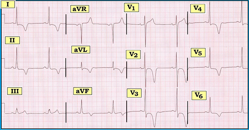
Scenario: No history is available for the ECG shown above. How would you interpret this tracing? What clinical conditions should be considered as the possible cause for the ST-T wave changes seen?
Interpretation: The rhythm is sinus bradycardia and arrhythmia, with an overall heart rate just under 60/minute. All intervals (PR/QRS/QT) are normal. The axis is normal at +50 degrees. Voltage for LVH is present (deepest S in V1, V2 + tallest R in V5, V6 ≥ 35 mm). With regard to Q-R-S-T changes, there are no Q waves and transition occurs early (with relatively tall R waves in leads V1, V2). The most remarkable finding on this tracing is diffuse, deep symmetric T wave inversion. T wave inversion almost attains a depth of 15 mm in leads V2, V3. Other subtle but real ST-T wave findings include 1-2 mm of J-point ST depression in multiple leads — suggestion of ST segment coving in leads I, aVL, V2, V3, V4 — and a hint of ST elevation in leads III, aVR, and V1.
The overall impression of this ECG is suggestive of Giant T wave syndrome. Although some T wave inversion is common in many conditions, the term "giant T waves" is reserved for a select number of clinical entities that produce truly deep (> 5 mm amplitude) T wave inversion. When this clinical picture is seen (as it is in the figure above), one should think of the following diagnostic entities: 1) apical (Yamaguchi) cardiomyopathy; 2) severe central nervous system (CNS) disorders (increased intracranial pressure); 3) Stokes-Adams attacks (especially when due to severe bradycardia or complete AV block); 4) acute ischemia/coronary artery disease; 5) post-tachycardia syndrome; and 6) massive pulmonary embolism (with acute right heart strain).
Without any history, it is impossible to know which of the above entities is most likely. Acute CNS disorders (stroke, subarachnoid or intracranial hemorrhage, seizure, coma, brain tumors, trauma) may produce some of the most bizarre ST-T wave abnormalities. However, the QT interval will usually be prolonged with CNS disorders and there will often be manifest T wave broadening (neither of which is seen here). Other stigmata of acute right heart strain usually accompany anterior T wave inversion from a large pulmonary embolus. The history is usually revealing when Stokes-Adams attacks, tachycardia, and/or CNS catastrophe is the reason for T wave inversion. This leaves us with myocardial ischemia and/or the special form of hypertrophic cardiomyopathy that preferentially affects the left ventricular apex. Increased QRS amplitude and the ST-T wave changes described above are consistent with either of these two entities.
For more information on Giant T waves, please visit: https://www.kg-ekgpress.com/ecg_-_giant_t_waves/.
Subscribe Now for Access
You have reached your article limit for the month. We hope you found our articles both enjoyable and insightful. For information on new subscriptions, product trials, alternative billing arrangements or group and site discounts please call 800-688-2421. We look forward to having you as a long-term member of the Relias Media community.