ECG Review: A Multi-Chambered Problem
ECG Review
A Multi-Chambered Problem
By Ken Grauer, MD
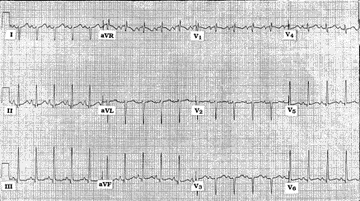 |
Figure. ECG obtained from a 60-year-old woman with shortness of breath.
Clinical Scenario: The 60-year-old woman whose ECG is shown in the Figure presented to the emergency department (ED) with shortness of breath. Can you guess why?
Interpretation: The rhythm is sinus tachycardia at a rate of 125 beats/min. The PR and QRS intervals are normal. The QT interval may be prolonged, although it is difficult to tell for sure given the rapid rate. Marked right axis deviation (RAD) is present, as determined by the predominantly negative QRS complex in lead I.
Regarding assessment for chamber enlargement (and the explanation of the title of this ECG Review, plus the answer to our clinical question)—we suspect that this patient has four-chamber enlargement. The tall peaked (pointed) P wave in standard lead II suggests right atrial enlargement (RAE). The notched P wave in lead I and the fairly deep (albeit pointed) negative component to the P wave in lead V1 suggests that there also may be left atrial enlargement (LAE). The surprisingly tall R wave in lead V6 (that exceeds 18 mm) is a less commonly invoked voltage criterion for left ventricular hypertrophy, but one that is probably accurate given the overall clinical picture. Finally, we suspect right ventricular hypertrophy (RVH). Admittedly, QRS morphology in standard leads I, II, and III is consistent with left posterior hemiblock. However, the constellation of RAD, RAE and an rSr’ pattern in lead V1 is better explained by proposing RVH.
Possible explanation for this patient’s dyspnea (in view of the ECG findings of sinus tachycardia and four-chamber enlargement) include heart failure from dilated congestive cardiomyopathy and/or pulmonary hypertension from pulmonary emboli or end-stage pulmonary disease. Despite predominant negativity in lead I of this tracing, the reasons we don’t suspect lead misplacement or dextrocardia here include: 1) that a small positive deflection (r wave) is seen in lead I of this tracing; 2) both the P wave and T wave are upright in lead I here; 3) an upright P wave is seen in lead II; and 4) there is normal R wave progression in the precordial leads.
Subscribe Now for Access
You have reached your article limit for the month. We hope you found our articles both enjoyable and insightful. For information on new subscriptions, product trials, alternative billing arrangements or group and site discounts please call 800-688-2421. We look forward to having you as a long-term member of the Relias Media community.