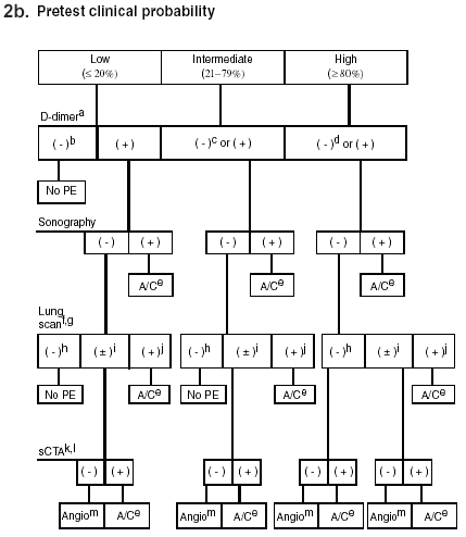
Fig. 2b: Suggested serial diagnostic testing strategy for patients presenting to the ED with varying clinical pretest probabilities of PE.
Key: a = conservative assessment of evidence supports use of VIDAS DD D-dimer assay; b = negative VIDAS DD D-dimer assay in low clinical pretest probability patients essentially excludes PE (< 1% posttest probability); c = evidence supporting use of negative VIDAS DD D-dimer result in intermediate clinical pretest probability patients for exclusion of PE appears valid; however, because the precision of the LR cannot be estimated, this test cannot be recommended at this time as unequivocally safe among the intermediate clinical probability patients; d = negative VIDAS DD D-dimer result does not exclude PE among high clinical pretest probability patients; e = anticoagulation; f = patients with underlying cardiopulmonary disease, especially chronic lung disease, may bypass radionuclide lung scanning and proceed directly to sCTA; g = ventilation scanning after a perfusion study, when indicated; h = negative lung scan finding indicates a reading of normal or near-normal; i = � finding on lung scan indicates a clinically indeterminate reading (i.e., neither normal/near-normal nor high probability [very low, low, intermediate, and so forth]); j = positive (+) lung scan indicates a high probability reading; k = sCTA is spiral (helical) computed tomography angiography with contrast, not requiring catheterization; l = a plausible alternative diagnosis seen on sCTA is currently the only evidence-based means of excluding PE with this test; m = conventional pulmonary angioplasty, requiring catheterization; PE = pulmonary embolism.
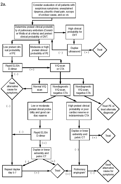
Figures used with permission from: (2a) Wolfe TR, Hartsell SC. Pulmonary embolism: Making sense of the diagnostic evaluation. Ann Emerg Med 2001;
37:509; and (2b) Gallagher EJ. Clots in the lung. Ann Emerg Med 2000;35:182.