Folic Acid and Neural Tube Defects
By Gerald T. Keegan, MD, FACS and Lynn Keegan, RN, PhD, HNC, FAAN
Neural tube defects (NTD) constitute an extensive spectrum of disease processes—from relatively mild manifestations to those that have a devastating impact on the individual sufferer, the extended family, and society as a whole. Myelomenigocele is the most common defect reported. The incidence of NTDs varies by geographic region, genetic or racial origin, and economic and educational status. Diseases (such as diabetes), medication history (especially antiepileptic medications), and occupational status play significant roles. The chances of a U.S. couple with no prior history of NTDs having a child with an NTD are approximately 1/500 to 1/1,000. Having a child with any type of NTD increases the chances of producing another child with a similar problem by 1/30, or about 3%. When two siblings are affected, the risk rises to as high as 12%.1 The incidence of the disease in the United States has declined over the years; part of this reduced incidence of NTDs may be related to intrauterine detection with subsequent elective termination of pregnancy.2
History of NTDs and Folic Acid
Epidemiological data incriminate environmental factors because the birth prevalence has varied over time and by geographical location; NTD epidemics appear to have taken place at different times and in different locations worldwide.3 During the 1970s and 1980s, there was an ongoing debate as to whether supplemental vitamins decreased the disease prevalence. This controversy was resolved by two randomized, multicenter studies. The first study found a 72% reduction in the recurrence of NTDs among at-risk women who received a 4 mg/d supplement of folic acid (FA) in the periconceptual period.4 The second study found that the risk of occurrence of first-time NTD was reduced by 60% through the use of periconceptual FA supplementation.5
Several additional randomized studies have demonstrated the critical role of FA supplementation in the prevention of NTDs. In 1992, the U.S. Public Health Service released its first recommendation of 0.4 mg/d FA consumption in all women of child-bearing age. Since that time, many national and international professional societies have released specific guidelines ranging from at least 0.4 mg/d for women of child-bearing age to 4 mg/d for women with a previous infant with NTDs. Since 1998, mandatory folate fortification of certain foods has been associated with at least a 54% reduction in the incidence of open NTDs in Canada. However, throughout the United States, Canada, and Europe, and more so in third world countries, the rates of periconceptual FA use remain suboptimal, especially among minorities.6
Lack of awareness and education regarding the benefits of folate is the most common reason women give for not choosing to use FA supplements before pregnancy. Only 25% of Canadian women meet the recommendation for women capable of becoming pregnant7 and educational programs have not resulted in increased supplement use.8 In Norway, researchers found that most pregnant women do not use FA as recommended, and a dramatically low usage was seen in immigrant populations.9 Despite educational programs in the Netherlands, a significant and persisting disparity in FA usage has been found in women of different socioeconomic status.10 Despite its ability to reach many U.S. women of child-bearing age with multiple messages about regularly using multivitamins, the Kaiser Health Plan found only a temporary small increase in usage by women who received repeated messages via mail.11 Health care providers must be alert to recognizing the more subtle NTD manifestations, such as hairy patches, dimples, fatty tumors of the lower back, and urinary tract symptoms.12 The findings suggestive of minimal NTDs in children will lead to the recommendation of higher doses of FA in those women at risk during subsequent pregnancies.
Clinical Presentation, Risk Factors, and Diagnosis
There are numerous presentations of NTDs. The most severe cases present with anencephaly, where there is an absence of major portions of the brain, scalp, and skull, and which is incompatible with life. The least severe cases present as spina bifida occulta in which only a bony defect is present. The manifestations of severe spina bifida may include infectious complications, paraplegia, bladder and bowel incontinence, Arnold-Chiari malformations, hydrocephalus, and, as a complication of hydrocephalus, diminished intelligence.13 Clinically, the most common problematic lesions include meningocoele with a meningeal sac but with intact neural elements; spina bifida cystica (myelomenigocele) with a skin-covered intact sac containing neural elements; and spina bifida aperta in which the sac is open.
Although most of these disorders are diagnosed at birth or in early childhood, an occasional abnormality, possibly mistaken for a skin or soft lesion indicating a minimal NTD, may be discovered on routine physical exam.12 The risk factors of giving birth to a child with an NTD include FA deficiencies, genetic abnormalities of FA metabolism, and insulin-dependent diabetes.14
The risk of women with epilepsy bearing children with congenital malformations is approximately twice that of the general population. The mechanism suspect in the production of NTDs is the use of antiepileptic medications that reduce the availability of maternal FA already diminished by pregnancy. Use of a higher FA dose (4 mg/d or equivalent) is protective.15 Valproate and carbamazine clearly have been incriminated in the development of NTDs and many of the newer anti-epileptic medications are suspect.14 Valproate-induced NTDs in mouse embryos have been ameliorated by substantially elevated and maintained levels of FA and vitamin B12 throughout the period of organogenesis.16 Studies demonstrating that phenytoin selectively inhibits neural tube closure also have shown that in early chick embryos FA decreased the phenytoin-induced NTD.17 Additional factors, including occupational exposure to teratogens, medications such as cyclophosphamide,18 excessive pregnancy weight, the use of oral contraceptives, and other nutrient deficiencies, can be contributory.14
Pathogenesis of NTDs
NTDs arise during the first few weeks after conception. The defect may begin developing in the unborn baby before the mother even knows she is pregnant. Many factors are involved in the proper closure of the neural tube. In human embryogenesis, the malformation generally is created by abnormal neural tube closure between the third and fourth weeks of gestational age. There is evidence, at least in other mammals, that the closure of the neural tube is intermittent at four discrete locations. Disruption of the process of fusion at any of these four sites may lead to an NTD, possibly arising through the mediation of site-specific genetic mechanisms. The genes responsible for this process include those of the folate metabolic pathway as well as those involved in folate transport.19 Studies in FA-binding protein-deficient mice have shown that the deficiency of this factor impacted the expression of several downstream signaling molecules and contributed to failure of neural tube closure and the development of craniofacial defects.20
Another interesting ingredient in the pathogenesis of NTDs is the relationship with homocysteine. This amino acid is involved in several key metabolic pathways including methylation and sulphuration. Blood concentrations of this substance are moderated by various dietary factors, including FA and vitamin B12, as well as by alterations in physiology, including renal impairment, and by variation in the activity of enzymes as a result of genetic polymorphism. In normal pregnancy, homocysteine levels should fall. Disturbances of this normal mechanism under atypical genetic control may produce metabolic abnormalities in both the mother and the fetus, resulting in elevated homocysteine levels associated with NTDs.21
In vitro studies of the behavior of neuroepithelial, cranial, and cardiac neural crest cells showed that the addition of FA increased neuroepithelial cell outgrowth and increased neural crest cell differentiation into nerve and smooth muscle cells. The addition of homocysteine to the cell culture increased the neural cell outgrowth but inhibited neural crest cell differentiation.22 The authors concluded that NTDs caused by folate deficiency and hypercysteinemia developed secondary to increased neuroepithelial to neural crest transformation, leading to a shortage of neuroepithelial cells in the neural tube. The consistent findings that effective FA supplementation prevents the defect and that high homocysteine levels always are present in the mothers of children with NTDs have led to extensive studies of the genes involved in FA and homocysteine metabolism. A comprehensive list of genes known to be involved in folate and homocysteine metabolism has been identified.23 Some of the more important studies have involved genes encoding the enzymes methylenetetrahydrofolate reductase or methylenetetrahydrofolate dehydrogenase (MTHFD) in FA metabolism. Genes encoding methionine synthase, its regulator methinine synthase reductase, and cystathionine synthase in homocysteine metabolism are also being studied.23,24 Studies in Ireland (a country with an historically high rate of NTDs) have shown that genetic variation in the MTHFD1 gene is associated with an increase in the genetically determined risk that a woman will bear a child with NTDs. This gene also may be associated with decreased embryo survival.25
Another recent study of five common functional polymorphisms in enzymes involved in homocysteine metabolism in young adults found that serum folate, red cell folate, vitamin B12, and total plasma homocysteine concentration (tHcy) were influenced significantly by the variant genotype MTHFR 677C>T. A particularly strong interaction was observed between MTHFR 677TT genotype and FA, which led to a high tHcy phenotype that was more pronounced in males. The genetic contribution to the variance was estimated at only 9%, with approximately 35% being attributed to low folate and vitamin B12 levels. This study emphasizes that dietary factors were most important in the control of the tHcy levels in young adults with additional somewhat weaker genetic effects, and further stresses the importance of the implementation of folate/B-vitamin food fortification programs.26 Many studies in laboratory animals have demonstrated that even in the presence of known genetic defects, FA can prevent the genetic expression of NTDs.27
FA Sources and Metabolism
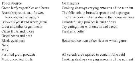
FA is a B-vitamin obtained primarily from yeasts, leafy vegetables, and animal liver. (See Table.) The vitamin cannot be synthesized in animals and requires intake in the diet. Since 1998, the U.S. Food and Drug Administration has mandated that cereal products be fortified with 140 mcg of FA per 100 g. It is important to be able to adequately assay that content, and a microbiological trienzyme extraction procedure has been developed for that purpose. Use of these techniques has demonstrated differences in FA content in lentils and peas derived from different locations.28 The protective effects of FA may be dose-related. Data on vitamin intake from more than 23,000 women from the northeastern United States were submitted to multiple logistic regression and restricted spline regression modeling with the finding that for each additional 500 dietary folate equivalents consumed per day, the prevalence of NTDs decreased by 0.78 cases. This study concluded that total folate dose, rather than supplemental folate alone, should be considered in formulating public health guidelines for NTD prevention.29 When ingested or stored in the liver, FA exits as a polyglutamate form. By a process of removing some of the glutamate residues, the intestinal cell lysosomes make the charge of the folate more positive and capable of absorption through the basal membrane of the epithelial cells and into the blood stream. Within the liver, the FA is reduced to a tetrahydrofolate (THF) through the action of dihydrofolate reductase, an NADPH-requiring enzyme. The function of THF is to carry and transfer various forms of one-carbon units during biosynthetic reactions. The role of vitamin B12 and the one carbon-THF’s in the conversion of homocysteine to methionine can have a significant impact on the availability of the active form of FA to the cells.30
|
Precautions in the Use of FA
FA is a water-soluble vitamin and it is unlikely that toxic levels could be reached under normal circumstances including standard vitamin and food supplementation. Doses in excess of 20 g/d for an extended period can produce renal damage. Concern has been raised as to whether excessive consumption of enriched cereal—in the face of significantly higher assayed levels of FA than listed on the food label—could lead to iron and FA toxicity, but considering the very high levels of FA needed for toxicity, this seems unlikely.31 However, the potential exists that FA supplementation could disguise underlying B12 deficiency, which itself has the potential to produce NTDs.32 Synthetic L-5-methyltetrahydrofolate has been proposed as being more appropriate as a fortificant because it is unlikely to mask the hematologic indicators of B12 deficiency.33 Potential dangers exist that with supplementation of dietary staples, such as flour, with FA as opposed to the natural folate (N5CH3HFGlu1), the FA could enter the cell and the metabolic pathway by a cobalamin (B12)-independent pathway and exacerbate or perhaps even induce B12 deficiency.34 In addition, there is a potential interaction between zinc plasma levels and FA metabolism as well as evidence suggesting a relationship between NTDs and lower levels of zinc as measured in the hair. For this reason all supplements should contain both zinc and folic acid.35,36
Recommendation
Folic acid supplementation is highly recommended in all women of child-bearing age in a dosage of at least 0.4 mg/d. Women who are at risk because of diabetes, insulin-dependent diabetes, and having other children with even minimal NTDs should take at least 4 mg/d because of the clear protective effects both in the genetic and acquired forms of NTD. Compliance issues have been of concern and might be improved by weekly rather than daily usage. Studies in Mexico demonstrated that there was a 50% decrease in the incidence of NTDs using a single tablet of 5 mg FA weekly as an alternative to daily supplementation.37 Investigators in New Zealand have shown a weekly dose of 2.8 mg FA was as effective as daily doses of 0.4 mg in lowering homocysteine levels in normal women of childbearing age.38 Educational programs and physician counseling regarding the benefits of FA are essential.
Gerald T. Keegan, MD is Emeritus Staff, Scott & White Clinic and Hospital, and former Professor of Surgery (Urology), Texas A&M University School of Medicine.
References
1. Toriello HV. Periconceptual vitamin supplementation for prevention of NTD: A review. Spina Bifida Ther 1982;4:603.
2. Frey L, Hauser WA. Epidemiology of neural tube defects. Epilepsia 2003;44(Suppl 3):4-13.
3. Oakley GP. Folic acid preventable spina bifida and anencephaly. JAMA 1993;269:1292-1293.
4. MRC Vitamin Study Research Group: Prevention of neural tube defects: Results of the Medical Research Council Vitamin Study. Lancet 1991;338:131-137.
5. Werler MM, et al. Periconceptual folic acid exposure and risk of occurrent neural tube defects. JAMA 1993;269:1257-1261.
6. McDonald SD, et al. The prevention of congenital anomalies with periconceptual folic acid supplementation. J Obstet Gynaecol Can 2003;25:115-121.
7. French MR, et al. Folate intakes and awareness of folate to prevent neural tube defects: A survey of women living in Vancouver, Canada. J Am Diet Assoc 2003;103:181-185.
8. Bailey LB, et al. Folic acid supplements and fortification affect the risk for neural tube defects, vascular disease and cancer: Evolving science. J Nutr 2003;133:1961S-1968S.
9. van der Pal-de Bruin KM, et al. Influence of educational level on determinants of folic acid use. Paediatr Perinat Epidemiol 2003;17:256-263.
10. Langley-Evans SC, Langley-Evans AJ. Use of folic acid supplements in the first trimester of pregnancy. J R Soc Health 2002;122:181-186.
11. Lawrence JM, et al. Design and evaluation of interventions promoting periconceptual multivitamin use. Am J Prev Med 2003;25:17-24.
12. Koester MC, Amundson CL. An unusual scalp lesion in a 15-year-old girl: A case report. J Athl Train 2001;36:182-184.
13. Hoffman HJ. Spinal dysraphism. Am Fam Physician 1987;36:129-136.
14. Yerby MS. Clinical care of pregnant women with epilepsy: Neural tube defects and folic acid supplementation. Epilepsia 2003;44(Suppl 3):33-40.
15. Weber M, Dib M. Folic acid and prevention of anomalies of foetal neural tube closing in women treated for epilepsy [in French]. Rev Neurol (Paris) 2003;159:165-170.
16. Padmanabhan R, Shafiullah MM. Amelioration of sodium valproate-induced neural tube defects in mouse fetuses by maternal folic acid supplementation during gestation. Congenit Anom Kyoto 2003;43:29-40.
17. Guney O, et al. The effects of folic acid in the prevention of neural tube development defects caused by phenytoin in early chick embryos. Spine 2003;28:442-445.
18. Ma A, et al. Effect of folic acid and supplemented with vitamin A and vitamin E on depressing teratogenesis induced by cyclophosphamide [in Chinese]. Wei Sheng Yan Jiu 2001;30:343-346.
19. Finnell RH, et al. Pathobiology and genetics of neural tube defects. Epilepsia 2003;44(Suppl 3):14-23.
20. Tang LS, Finnell RH. Neural and orofacial defects in Folbp1 knockout mice. Birth Defects Res Part A Clin Mol Teratol 2003;67:209-218.
21. Hague WM. Homocysteine and pregnancy. Best Pract Res Clin Obstet Gynaecol 2003;17:459-469.
22. Boot MJ, et al. Folic acid and homocysteine affect neural crest and neuroepithelial cell outgrowth and differentiation in vitro. Dev Dyn 2003;227:301-308.
23. Gos M Jr, Szpecht-Potocka A. Genetic basis of neural tube defects. II. Genes correlated with folate and methionine metabolism. J Appl Genet 2002;43:511-524.
24. Zhu H, et al. Homocysteine remethylation enzyme polymorphisms and increased risks for neural tube defects. Mol Genet Metab 2003;78:216-221.
25. Brody LC, et al. A polymorphism, R653Q, in the trifunctional enzyme methylenetetrahydrofolate dehydrogenase/methenyltetrahydrofolate cyclohydrolase/formyltetrahydrofolate synthetase is a maternal genetic risk factor for neural tube defects: Report of the Birth Defects Research Group. Am J Hum Genet 2002;71:1207-1215.
26. Kluijtmans LA, et al. Genetic and nutritional factors contributing to hyperhomocysteinemia in young adults. Blood 2003;101:2483-2488.
27. McLone DG. The etiology of neural tube defects: The role of folic acid. Childs Nerv Syst 2003;19:537-539.
28. Han JY, Tyler RT. Determination of folate concentrations in pulses by a microbiological method employing trienzyme extraction. J Agric Food Chem 2003;51:5315-5318.
29. Moore LL, et al. Folate intake and the risk of neural tube defects: An estimation of dose-response. Epidemiology 2003;14:200-205.
30. THE Medical Biochemistry Page. Introduction to Vitamins. Available at: www.indstate.edu/thcme/mwking/vitamins.html. Accessed Oct. 17, 2003.
31. Whittaker P, et al. Iron and folate in fortified cereals. J Am Coll Nutr 2001;20:247-254.
32. Suarez L, et al. Maternal serum B12 levels and risk for neural tube defects in a Texas-Mexico border population. Ann Epidemiol 2003;13:81-88.
33. Venn BJ, et al. Comparison of the effect of low-dose supplementation with L-5-methyltetrahydrofolate or folic acid on plasma homocysteine: A randomized placebo-controlled study. Am J Clin Nutr 2003;77:658-662.
34. Weir DG, Scott JM. Brain function in the elderly: Role of vitamin B12 and folate. Br Med Bull 1999;55:669-682.
35. Nogueira Ndo N, et al. Changes in plasma zinc and folic acid concentrations in pregnant adolescents submitted to different supplementation regimens [in Portuguese]. Cad Saude Publica 2003;19:155-160.
36. Srinivas M, et al. Association between lower hair zinc levels and neural tube defects. Indian J Pediatr 2001;68:519-522.
37. Martinez de Villarreal L, et al. Decline of neural tube defects cases after a folic acid campaign in Nuevo Leon, Mexico. Teratology 2002;66:249-256.
38. Adank C, et al. Weekly high-dose folic acid supplementation is effective in lowering serum homocysteine concentrations in women. Ann Nutr Metab 2003;47:55-59.
Keegan GT, Keegan L. Folic acid and neural tube defects. Altern Ther Women's Health 2003;5(11):81-85.
Subscribe Now for Access
You have reached your article limit for the month. We hope you found our articles both enjoyable and insightful. For information on new subscriptions, product trials, alternative billing arrangements or group and site discounts please call 800-688-2421. We look forward to having you as a long-term member of the Relias Media community.