Cardiac Biomarkers
Cardiac Biomarkers
Author: Donald H. Schreiber, MD, CM, FRCPC, FACEP, Associate Professor of Emergency Medicine, Stanford University School of Medicine, Stanford, CA.
Peer Reviewer: Amal Mattu, MD, Associate Professor and Program Director, Emergency Medicine Residency, University of Maryland School of Medicine, Baltimore, MD.
Introduction
An accurate, rapid biomarker of the acute coronary syndrome (ACS) is the current holy grail of emergency medicine. The goal is tantalizingly close. In the ED, the greatest challenge in decision making often is encountered when a low-risk patient presents with atypical chest pain, few risk factors, a nondiagnostic electrocardiogram, and normal baseline cardiac markers for myocardial necrosis. The evaluation of patients with dyspnea and possible congestive heart failure (CHF) also may be difficult. This issue reviews selected articles from the current literature on cardiac biomarkers and helps guide physicians in their use and interpretation.
Acute nSTEMI
Source: Fesmire FM, Decker WW, Diercks DB, et al. Clinical policy: critical issues in the evaluation and management of adult patients with non-ST-segment elevation acute coronary syndromes. Ann Emerg Med 2006;48:270-301
This article summarizes the American College of Emergency Physician's (ACEP's) clinical policy on the diagnosis and management of acute non-ST segment elevation acute myocardial infarction (nSTEMI).
ACEP provides three level B recommendations for ruling out nSTEMI in the ED:
1) A single negative CK-MB (creatine kinase myocardial band) mass, troponin-I or troponin-T measured 8-12 hours after symptom onset.
2) A negative myoglobin in conjunction with a negative CK-MB mass or negative troponin-I measured at baseline and 90 minutes in patients presenting less than eight hours after symptom onset.
3) A negative two-hour delta CK-MB in conjunction with a negative two-hour delta troponin-I in patients presenting less than eight hours after symptom onset.
In their paper, the ACEP authors review the evidence for a 90-minute rule out in the ED utilizing myoglobin in conjunction with either CK-MB or troponin-I. The CK-MB/myoglobin protocol yielded a sensitivity of only 92% at 90 minutes. The myoglobin/troponin-I combination yielded a sensitivity of 97% at 90 minutes.
The ACEP authors cited problems with the relative lack of specificity for myoglobin and the fact that many studies were not based on the current internationally accepted definition of acute myocardial infarction (AMI) adopted by the American College of Cardiology (ACC) and the European Society of Cardiology (ESC).
According to the ACC and the ESC, AMI is defined as a typical rise and fall of biochemical markers (e.g., troponin and CK-MB), with at least one of the following:
- Ischemic symptoms,
- New pathologic Q waves on ECG,
- Ischemic ECG changes (ST-segment elevation or depression),
- Coronary artery intervention; or
- Pathologic findings of an AMI.
Commentary
The authors review the important issues related to troponin assays. In the laboratory, the various commercially available CK-MB mass immunoassays and myoglobin assays are fairly similar even though there are no international standardized reference ranges established. Troponin-T assays are only produced by a single manufacturer and no laboratory variability exists. However, troponin-I assays vary widely; there is up to a 20-fold variation among the commercially available troponin-I assays.
The clinician also must understand the concept of 99th percentile cutoff levels and 10% coefficient of variation (CV) cutoff levels for troponin-I. The 99th percentile level is the upper limit of normal in a healthy patient population and represents the most sensitive detection level for a particular assay. Every assay also has a percentage variation when the same aliquot of blood or serum is repeatedly tested, the coefficient of variation (CV), and is expressed as a percentage. A 10% CV indicates a cutoff level where this variation between sample runs is less than 10%. As the level of troponin-I in the blood sample falls closer to the lower limit of sensitivity, the CV rises dramatically. Therefore, the 10% CV level is always higher than the 99th percentile cutoff level. Each manufacturer reports these values in their package inserts.
Note that for most of the assays, the ratio of the CV level to the 99th percentile level is about 3:1. In certain clinical scenarios, patients may have troponin-I results intermediate to these two cutoffs. This laboratory finding is of uncertain clinical significance. It is imperative that the emergency physician understand that this detectable level of troponin-I may either be a true positive finding or a laboratory variation (false-positive elevation) but cannot be ignored. Serial determination of troponin-I will help elucidate the true nature of these intermediate results.
In the absence of standardized controls for troponin-I, the National Academy of Clinical Biochemistry has recommended that laboratories use the 10% CV level as the laboratory cutoff for troponin elevation. However, levels that are intermediate to the 10% CV cutoff and the 99th percentile level also are associated with increased risk of adverse cardiac events. Ultimately, the 99th percentile level may become the recommended cutoff despite the higher rate of false positives for the ACS.
It is difficult to comprehend the ACEP clinical policy committee recommendations on the 90-minute rule out that accepts a missed AMI rate of 3-8%.
ACEP's final recommendations on the utilization of delta CK-MB and delta troponin-I are based on one of the authors' own work. A Delta troponin-I evaluation as recommended by Fesmire is partially based on his scientific work utilizing older second generation troponin-I assays. Fesmire only evaluated a second generation troponin-I assay, the Stratus CS assay, on a small study of 125 patients. No data were reported on how many troponin I samples were elevated at baseline or how many were elevated at the two-hour time point. Results below the analytical sensitivity of the assay were artificially rounded up to the threshold level and then used in the calculations. The threshold levels used in the original study (0.03 ng/mL) were different than those cited in the current article for the 99th percentile level (0.06 ng/mL) and the 10% CV level (0.07 ng/mL). The authors only reported AUC (area under the curve) calculations without mentioning sensitivity. The criteria for AMI also was based on a higher 2.0 ng/mL AMI cutoff on the main laboratory instrument using older WHO criteria.
ACEP's recommendations for delta troponin-I, therefore, appears to be based on very limited data utilizing only one commercial assay and on other small studies with significant methodological limitations. ACEP's recommendations may not be generalizeable to other commercially available assays.
A potential value for delta troponin-I may be in the clinical setting where both results are intermediate to the 99th percentile level and the 10% CV level on specific laboratory platforms.
Overall, ACEP's committee does stress the importance of serial measurements over 8-12 hours after symptom onset to rule out AMI. Emergency physicians also must recognize that ACS associated with unstable angina without infarction will not show a rise in these markers of myocardial necrosis. These cardiac markers cannot be utilized to exclude myocardial ischemia.
Caution must, therefore, be used when evaluating these recommendations for serial marker determination from ACEP in ED patients with suspected ACS.
Cardiac Biomarkers for Acute Coronary Syndromes
Source: Storrow AB, Lindsell CJ, Han JH, et al. Discordant cardiac biomarkers: frequency and outcomes in emergency department patients with chest pain. Ann Emerg Med 2006;48:660-665.
Based on a secondary analysis of the prospective multicenter Internet Tracking Registry of Acute Coronary Syndrome, 8769 eligible ED patients out of a total of 17,713 were identified who met their inclusion criteria. Patients were diagnosed with ACS based on events (death or myocardial infarction), revascularization (PCI, CABG), positive stress test results (nuclear or other stress test for ischemia), or cardiac catheterization findings of > 70% stenosis in any coronary vessel. Patients were excluded for repeat ED visits, STEMI, if no ECG diagnostic category was recorded; or if no CK, CK-MB, and troponin I were obtained within one hour of each other.
Of the patients, 18.4% had ACS. Discordant CK-MB and troponin results were present at baseline in 7% (CK-MB+/troponin- in 4.9%; CK-MB-/troponin+ in 2.1%). Elevated CK-MB (CK-MB+) with negative total CK (CK-) levels occurred in 3.1%. The unadjusted odds ratios (ORs) for ACS with discordant markers were as follows: CK-MB+/CK- 5.7 (4.4-7.4); CK-MB+/CK+ 4.4 (3.6-5.2); CK-MB-/Tn+ 4.8 (3.4-6.8); CK-MB+/Tn- 2.2 (1.7-2.8); CK-MB+/Tn+ 26.6 (18.0-39.3).
The authors concluded that an elevated troponin level, regardless of CK-MB level, and an elevated CK-MB level, regardless of troponin level, identify patients at higher risk of ACS.
The authors recommend a multimarker strategy incorporating CK-MB and troponin to identify patients with suspected ACS in the ED.
Commentary
Caution must be used when interpreting the authors' recommendations; older second-generation troponin assay platforms were used. Some centers used troponin-I and others troponin-T. The degree of discordant troponin and CK-MB results may be different with the current third generation troponin-I and troponin-T assays.
Only baseline marker results were used in this study and discordance also may be reflected by variations in the time from patient symptom onset to the blood collection time in the study. Time intervals were not reported by the authors.
In their analysis of the CK-MB+/CK- subgroup, the authors did not report the number of patients with a positive troponin result. A better analysis might have focused on the CK-MB+/CK-/Tn- subgroup. The higher odds ratio (OR) in the CK-MB+/CK- subgroup might be primarily related to the number of positive troponin patients rather than the discordant CK-MB/CK results.
Despite their conclusions on the prognostic value of CK-MB, the CK-MB+/Tn- group had an unadjusted OR of only 2.2 versus the CK-MB-/Tn+ subgroup with 4.8. Their findings actually support other reports that CK-MB has less prognostic value than troponin.
The authors recommend a multimarker strategy incorporating CK-MB and troponin despite other reports that suggest an isolated CK-MB has limited prognostic value and other studies that call for the establishment of a single marker strategy utilizing troponin alone.
The practicing emergency physician must still be cognizant of the fact that negative baseline markers analyzed within six hours of symptom onset do not rule out AMI with sufficient sensitivity.
Elevated Cardiac Troponin Levels
Source: Jeremias A, Gibson CM. Narrative review: alternative causes for elevated cardiac troponin levels when acute coronary syndromes are excluded. Ann Intern Med 2005;142:786-791.
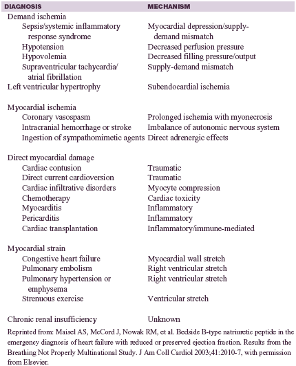
The authors in this cogent review discuss the many other clinical scenarios that lead to the elevation of cardiac troponin in the absence of thrombotic ACS and epicardial coronary artery disease (CAD).
They cited evidence to illustrate that troponin may be elevated in the setting of transient myocardial ischemia. The notion that troponin is only increased after irreversible myocardial necrosis has been challenged by the observation of transient elevations in the setting of tachyarrhythmias and Prinzmetal's angina. In patients with septic shock and the systemic inflammatory response syndrome, elevated cardiac troponins are common but largely occur in patients without significant CAD. Demand ischemia has been a proposed mechanism that explains this observation. Demand ischemia occurs when there is a significant mismatch between myocardial oxygen demand and supply in the absence of flow limiting CAD. Hypotension and hypovolemic shock, as well as tachyarrhythmias, may lead to increased cardiac troponin via demand ischemia.
Table 1 outlines other scenarios that are associated with elevated cardiac troponin in the absence of CAD.
| Table 1. Nonthrombotic causes and presumed mechanism for elevated cardiac troponin level |
|
Commentary
For the emergency physician, it is important to recognize that a single elevation of cardiac troponin may not be caused by ACS. In a recent study of 1000 patients, troponin I was elevated on admission in 11.2% of patients, but 45% of these patients had a final diagnosis other than ACS. The conditions listed in Table 1 are useful for elucidating a differential diagnosis of troponin elevation.
Bedside B-Type Natriuretic Peptide in Heart Failure
Source: Maisel AS, McCord J, Nowak RM, et al. Bedside B-Type natriuretic peptide in the emergency diagnosis of heart failure with reduced or preserved ejection fraction. Results from the Breathing Not Properly Multinational Study. J Am Coll Cardiol 2003;41:2010-2017.
The Breathing Not Properly multicenter prospective study involved 1586 patients presenting to the ED with acute dyspnea. Patients younger than 18 years and those receiving dialysis were excluded. BNP (B-type natriuretic peptide) levels were measured with a rapid bedside assay. Independent cardiologists who were blinded to the BNP results clinically diagnosed CHF as the ultimate etiology of the patients' dyspnea.
In the study cohort, 47% of patients were ultimately diagnosed with dyspnea secondary to CHF, 49% did not have CHF, and 4% had a history of LV (left ventricle) dysfunction but dyspnea was attributed to other causes. BNP levels had significant predictive power for the diagnosis of CHF exceeding the predictive power of several other common clinical variables of CHF. Cardiomegaly on chest radiography, a history of CHF, rales on physical examination, and a history of paroxysmal nocturnal dyspnea had CHF diagnostic accuracies of only 81%, 75%, 69%, and 60%, respectively. BNP, with a cutoff value of 150 pg/mL, had a diagnostic accuracy of 84% (sensitivity, 85%; specificity, 83%), and an AUC of 0.91. Given the potentially severe consequences of delayed or missed diagnosis of CHF, the authors proposed a conservative BNP cutoff level of 100 pg/mL to rule out CHF as a cause of dyspnea in the ED. Applied to their patients, this cutoff provided a NPV (negative predictive value) of 89% (sensitivity, 90%; specificity, 76%).
A subset of 452 patients from this study with a final diagnosis of CHF underwent echocardiography within 30 days of enrollment. Of the 452 patients with CHF, 37% had diastolic failure, whereas 64% had systolic failure. BNP levels were lower in patients with diastolic failure than in those with systolic failure (median, 413 versus 821 pg/mL). The BNP cutoff of 100 pg/mL had a sensitivity of 86% and an NPV of 96%. Using multiple logistic regression, BNP was the single strongest predictor of systolic dysfunction versus diastolic dysfunction, followed by oxygen saturation, history of myocardial infarction (MI), and heart rate. The authors recognized that the higher the BNP level, the more likely it was that the patient had systolic dysfunction.
The authors also concluded that a BNP cutoff of > 500 pg/mL had a positive predictive value (PPV) of 90% for the diagnosis of CHF. Higher values also correlated with an increased risk of morbidity and mortality.
Commentary
In the Breathing Not Properly study, the attending emergency physician's impression of CHF as the etiology of dyspnea also was evaluated. When the physician estimated probability for CHF was > 80%, clinical judgment had a sensitivity of only 49% but a specificity of 96% compared to the 90% sensitivity and 76% specificity for a BNP level > 100 pg/mL. However, in 43% of patients, the physician's impression was intermediate (20-80% probability of CHF) but the BNP level correctly diagnosed CHF in 74% of these patients. Overall, the AUC was 0.86 (95% confidence interval [CI] 0.84-0.88) for clinical judgment alone and 0.90 (95% CI 0.88-0.91) for BNP alone using the 100 pg/mL cutoff. The combination of clinical judgment and BNP achieved an AUC of 0.93 (95% CI 0.92-0.94). This result should not be surprising given the complimentary sensitivity and specificity of clinical judgment and objective testing.
The recommended thresholds of < 100 pg/mL to rule out CHF and > 500 pg/mL to rule in CHF have been calculated to yield a negative likelihood ratio (LR) of 0.13 and a positive LR of 8.1. (Random chance has an LR of 1.0 by definition.) Intermediate values of BNP between 100-500 pg/mL have a positive LR of only 1.9. Therefore, an intermediate BNP result alone may not be used to rule in or rule out CHF.
To explore the value of clinical variables in conjunction with an intermediate BNP value, the Breathing Not Properly group calculated their positive likelihood. The clinical variables included cephalization of the pulmonary vasculature on chest x-ray, a history of CHF or CAD, lower-extremity edema, pulmonary edema, cephalization of the pulmonary vasculature, or cardiomegaly. The LR+ for a history of CHF alone was 4.6. An intermediate BNP level together with a history of CHF had a modest cumulative LR+ of 4.3; this was not significantly different than the LR+ for a history of CHF alone. However, any combination of two or more of the above clinical features, a history of CHF, and a mid-range BNP level had a cumulative LR+ of 10. A mid-range BNP level, without a history of CHF, and none or only one of these clinical criteria does not rule out CHF and only had a cumulative LR- of 0.7. Intermediate results must be interpreted with caution and by themselves do not rule in or out the diagnosis of CHF.
Dyspnea in the ED
Source: Januzzi JL Jr, Camargo CA, Anwaruddin S, et al. The N-terminal Pro-BNP investigation of dyspnea in the emergency department (PRIDE) study. Am J Cardiol 2005;95:948-954.
Analogous to the Breathing Not Properly study for BNP, the ProBNP Investigation of Dyspnea in the Emergency Department (PRIDE) study established cutoff values for NT-proBNP. This study had two goals: 1) to set clinically useful cutoff points for NT-proBNP as an aid to the diagnosis of CHF; and 2) to compare NT-proBNP levels with the clinical probability assessment of acute CHF by the attending emergency physician. The investigators prospectively examined 600 patients who presented to the ED with dyspnea, excluding patients younger than age 21 and those with ischemic ECG changes. They evaluated NT-proBNP levels. The diagnosis of CHF was independently confirmed by a panel of cardiologists who were blinded to the NT-proBNP results.
Overall, 35% of patients had an ultimate diagnosis of CHF exacerbation; their median NT-proBNP value was 4054 pg/mL. For patients with a history of CHF but whose dyspnea was attributed to another cause, the median level was 1175 pg/mL. For patients without CHF, the median was 114 pg/mL. Overall, NT-proBNP testing generated an AUC of 0.94 with a NT-proBNP cutoff of 900 pg/mL.
The investigators observed an age-related variation in the sensitivity and specificity of NT-proBNP for ruling in CHF. For patients younger than age 50, the sensitivity of an NT-proBNP result > 900 pg/mL for ruling in CHF was only 73% (specificity, 96%), but it was 91% sensitive and 80% specific for patients ages 50 or older. This strong effect of age on the NT-proBNP level was likely due to age-related decreases in renal function.
Given this finding, the authors established separate cutoff values based on age. Age-specific thresholds to rule in CHF were 450 pg/mL for patients younger than age 50 and > 900 pg/mL for patients ages 50 or older. These thresholds were highly sensitive (91% and 93%, respectively) and specific (95% and 80%, respectively) for the diagnosis of acute CHF. The assay manufacturer proposed a third age-related rule-in cutoff of 1800 pg/mL for patients older than age 75.
Commentary
All three cutoff points were prospectively evaluated in a subsequent study of 1256 patients with dyspnea. Utilizing the three age-dependent NT-proBNP rule-in levels, an overall sensitivity of 90% and a specificity of 84% were achieved for the diagnosis of acute CHF. In summary, patients with shortness of breath and NT-proBNP levels above the age-dependent thresholds of 450 pg/mL for those younger than age 50, 900 pg/mL for patients ages 50-75, and 1800 pg/mL for patients older than age 75 were most likely to have acute CHF.
With respect to ruling out CHF, the investigators proposed a single age-independent threshold of 300 pg/mL that yielded a sensitivity of 99% and an NPV of 99% when applied to all patients in the original derivative study cohort. In the subsequent follow-up study, the utility and accuracy of the single age-independent threshold of 300 pg/mL for ruling out CHF was confirmed with an NPV of 98%.
Multivariate analysis of the data revealed that, when age-stratified cutoff points were used, an increased NT-proBNP was the single strongest predictor of acute CHF compared to other clinical variables, such as interstitial edema on chest radiographs, orthopnea, use of loop diuretics before presentation, and rales on physical examination. Clinical findings of cough and fever were independently predictive of diagnoses other than CHF.
The emergency physician's clinical judgment was recorded on a scale of 0-100 to score the physician's estimated pre-test probability that the etiology of dyspnea was CHF. (The physicians were blinded to the NT-proBNP level.) Clinical judgment alone had an AUC of 0.90 that was statistically inferior to the AUC for the NT-proBNP level alone but similar to the AUC of 0.86 for clinical judgment alone in the multicenter Breathing Not Properly Study. Adding NT-proBNP results to the clinician's estimate further improved the sensitivity and specificity, leading to an AUC of 0.96, which was significantly better than that of either modality alone.
Shortness of Breath and B-Type Natriuretic Peptide
Source: Maisel A, Hollander JE, Guss D, et al. Primary results of the Rapid Emergency Department Heart Failure Outpatient Trial (REDHOT). A multicenter study of B-type natriuretic peptide levels, emergency department decision making, and outcomes in patients presenting with shortness of breath. J Am Coll Cardiol 2004;44:1328-1333.
The Breathing Not Properly investigators undertook the multicenter Rapid Emergency Department Heart Failure Outpatient Trial (REDHOT) to examine the prognostic role of BNP in the ED. They examined baseline BNP levels in 464 patients with dyspnea when they presented to the ED and evaluated its relationship with clinical decision making and clinical outcomes. Treating physicians were blinded to the BNP levels throughout the study. The primary outcome measure was the decision to admit or discharge the patient from the ED. A secondary composite 90-day outcome was based on cardiac mortality, subsequent ED visits, and rehospitalization for acute CHF.
About 90% of patients were admitted. Of interest, baseline BNP levels did not significantly differ between patients who were discharged and those who were admitted. Using logistic regression analysis, the decision to admit or discharge a patient and the initial severity of CHF based on the New York Heart Association (NYHA) classification score had no influence on 90-day outcomes. The BNP level was a strong predictor of the 90-day outcome. Of admitted patients, 11% had BNP levels < 200 pg/mL; however, the treating ED physician determined that 66% of these patients had disease of NYHA (New York Heart Association) functional class III or IV and that they were at risk for adverse outcomes. The 90-day composite adverse event rate in admitted patients with BNP < 200 pg/mL was 9%, significantly lower than the 29% rate of those admitted with BNP > 200 pg/mL. Overall, 26% of all admitted patients had an adverse outcome at 90 days versus 42% of patients who were discharged. Mortality rates at 90 days between the admitted and discharged groups did not significantly differ.
Commentary
This study highlighted the lack of correlation between the physician's clinical impression and the BNP level as an objective measure of disease severity. With the high cost of hospitalization and prolonged lengths of stay for CHF patients, the utility of BNP as a triage tool to guide admission decisions merits further study.
Ischemia-Modified Albumin in Diagnosing Myocardial Ischemia
Source: Anwaruddin S, Januzzi JL Jr, Baggish AL, et al. Ischemia-modified albumin improves the usefulness of standard cardiac biomarkers for the diagnosis of myocardial ischemia in the emergency department setting. Am J Clin Pathol 2005;123:140-145.
The authors studied 193 patients with symptoms suggestive of ACS presenting to a university teaching hospital; a majority of those patients were admitted. The studied evaluated the performance of IMA (ischemia-modified albumin) in conjunction with myoglobin, CK-MB, and troponin-I at baseline. Their analysis focused only on the 25 patients in the cohort who were subsequently diagnosed with ACS. They reported a high sensitivity of 80% and a poor specificity of only 31% but a high negative predictive value (NPV) of 92% when a cutoff value of 90 U/mL was utilized. A multimarker strategy that incorporated IMA with myoglobin, troponin I, and CK-MB yielded an NPV that approached 97%.
Commentary
Early investigations noted the increase in IMA levels in the setting of ischemia. A number of small studies have evaluated its use in ED patients with chest pain.
Many of these studies, including the one cited here, are limited by selection bias. The authors' analysis focused only on patients with a 100% prevalence of ACS instead of the more heterogeneous ED patient population with chest pain where the prevalence of ACS is much lower.
In other studies, IMA was found to be elevated in more than 70% of all patients with chest pain. The low specificity of the IMA assay is attributed to the false elevations that occur in liver disease, cancer, chronic renal insufficiency, CVA (cerebrovascular accident), and infections. There also is some overlap of values between normal healthy controls and patients with abnormal levels. Raising the cut-off level would improve specificity but at the expense of the negative predictive value.
The IMA test is currently FDA approved for low-moderate risk patients with negative markers of myocardial necrosis and a nondiagnostic ECG.
Further clinical evidence is required, especially in the generic heterogeneous ED patient population with chest pain, before this test can be generally recommended.
Myeloperoxidase in ACS
Source: Brennan ML, Penn MS, Van Lente F, et al. Prognostic value of myeloperoxidase in patients with chest pain. N Engl J Med 2003;349:1595-1604.
Myeloperoxidase (MPO) is a marker of oxidative stress and inflammation. Its role in the diagnosis and risk stratification of ACS was evaluated in this study of 604 ED patients with chest pain. MPO levels at presentation were higher in patients with an ultimate diagnosis of acute myocardial infarction than in those who did not have the diagnosis. In patients with a negative troponin-T at baseline, myeloperoxidase levels were significantly elevated in those patients who subsequently evolved an elevated troponin T than in those patients who were consistently negative for troponin T. MPO levels also predicted the risk of major adverse cardiac events at 30 days and at six months. MPO, in contrast to troponin T and CK-MB, identified patients at higher risk even in the absence of myocardial necrosis.
Commentary
As a result of this study, MPO has been marketed as a useful marker in the ED for risk stratification among ED patients with chest pain. However, this study was conducted on a relatively high-risk group of ED patients with chest pain. Of the patients, 23.5% were ultimately diagnosed with myocardial infarction, 17.1% had unstable angina, and only 21.5% were diagnosed with non-cardiac chest pain. MPO is a marker of neutrophil activation and is more sensitive to inflammatory changes than those induced by ischemia. It is unlikely that MPO would be a specific marker of myocardial ischemia and may be elevated in other diseases associated with inflammation or infection. Its performance in a more heterogeneous ED patient population with chest pain remains to be determined. However, as a marker of vulnerable coronary artery plaque, its value in risk stratification warrants further study. Assay standardization, establishment of reporting units, and the validation of imprecision and detection limits also are required.
Conclusions
The use of cardiac markers continues to evolve as new biomarkers become available and troponin assays become increasingly sensitive. The primary markers of choice continue to be the cardiac troponins T and I. The traditional CK-MB isoenzyme, although somewhat less sensitive and less specific than the troponins, continues to be used in most EDs. Myoglobin is an early marker of myocardial infarction but its lack of specificity limits its use. Serial determination of myoglobin improves sensitivity but still does not approach 100% for the detection of AMI. Negative troponin and CK-MB results within six hours of symptom onset are not sensitive enough to rule out acute myocardial infarction. Serial measurement of troponin and CK-MB through 6-12 hours after symptom onset is required. The availability of rapid, sensitive point-of-care troponin and CK-MB assays has the potential to speed patient throughput in the ED and facilitate serial measurement of cardiac markers. The introduction of beta natriuretic peptide assays and their use is reviewed extensively as studies continue to show that elevated levels are the single best predictor for the diagnosis of CHF in ED patients with dyspnea. New cardiac markers such as IMA and MPO are of limited use until further evaluation has taken place.
An accurate, rapid biomarker of the acute coronary syndrome (ACS) is the current holy grail of emergency medicine.Subscribe Now for Access
You have reached your article limit for the month. We hope you found our articles both enjoyable and insightful. For information on new subscriptions, product trials, alternative billing arrangements or group and site discounts please call 800-688-2421. We look forward to having you as a long-term member of the Relias Media community.