Critical Path Network: ACI: Update protocols or risk poor outcomes
Critical Path Network
ACI: Update protocols or risk poor outcomes
New approaches will ID more patients with MI
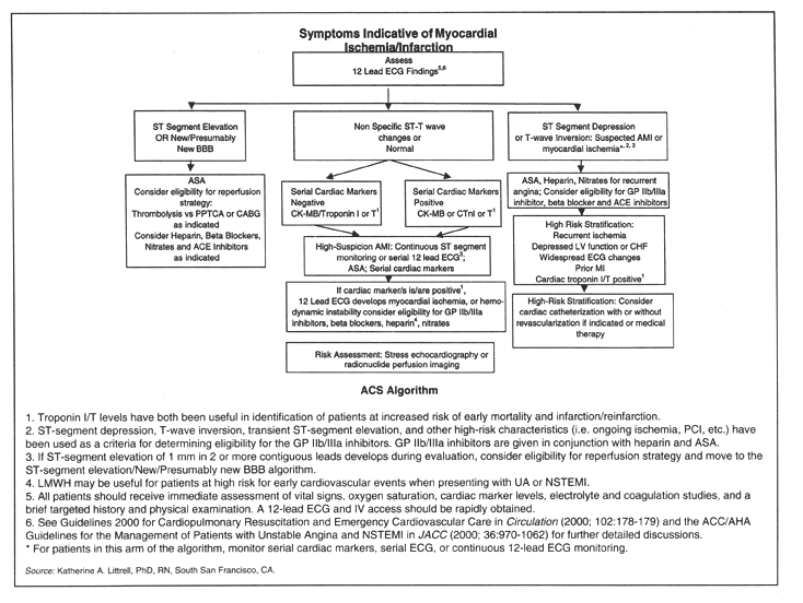
A patient comes to the emergency department (ED) with moderate chest pain and is discharged with a diagnosis of indigestion. Hours later, the patient dies of an undiscovered acute myocardial infarction (AMI).
Does this sound like your worst nightmare? Between 2% and 4% of ED patients who actually have an AMI are mistakenly sent home, warns Katherine A. Littrell, PhD, RN, project manager for the National Registry of Myocardial Infarction at Genentech, based in South San Francisco, CA.1
A new report from the Bethesda, MD-based National Heart Attack Alert Program (NHAAP), Evaluation of Technologies for Identifying Acute Cardiac Ischemia [ACI] in Emergency Departments, will help to ensure that patients don’t fall through the cracks, says Littrell.
There is a danger of misdiagnosis because ACI patients often have confusing and misleading symptoms, such as a normal or nondiagnostic 12-lead electrocardiogram (ECG), no chest pain or shortness of breath, and initially normal cardiac marker profiles, adds Littrell.
Update your protocols
Even though typical symptoms are often absent, these patients may actually have unstable angina, non-ST-segment elevation myocardial infarction, or ST-segment elevation myocardial infarction, she explains.
"These new recommendations will help you identify more patients with [ACI]," Littrell points out.
Use the report to update your protocols immediately, urges Julie Bracken, RN, MS, CEN, director of nursing education for the ED at Cook County Hospital in Chicago and the Des Plaines, IL-based Emergency Nurses Association representative to the NHAAP.
"ED nurses need to adapt their clinical approach to diagnostic technologies based on the updated technology report," Bracken insists.
You play an important role in influencing which diagnostic technologies are given to each specific patient presenting with actual or potential ACI, says Bracken.
"In this cost-conscious health care environment, guides to help direct proven technologies to diagnose difficult patients and expedite appropriate care are valued," she says.
Collaboration with physicians is important
Collaborate with physicians constantly to determine the best clinical approach for care, based on an individual patient’s response to technology, she advises.
Here are key recommendations of the report:
- Administer serial ECGs to patients with nondiagnostic 12-lead ECGs and symptoms of ACI.
A single ECG is not enough to rule in or rule out ACI, says Littrell.
Instead, patients with symptoms of ACI should receive serial ECGs or continuous ST-segment monitoring after arriving at the hospital, she advises. Research shows that 6.7% of these patients developed ST-segment elevation after their arrival, in a median time of 63 minutes, she notes.
"It appears that ST-segments are unstable in those early hours," says Littrell. "So serial ECGs are essential for patients with evolving ST-segment elevations," she emphasizes.2
- Use specific diagnostic tests only after general tests fail to diagnose ACI.
Broad use of such technologies as ECG and the Acute Ischemia Time-Insensitive Predictive Instrument (ACI-TIPI) for initial evaluation of all patients presenting with signs and symptoms of ACI is indicated, says Bracken.
"The results of these tests rule in the more high-risk cases for care," she adds.
For more challenging cases, you may need to use other testing to further evaluate ACI, says Bracken. To stay current with the report’s recommendations, change your protocols to include additional testing only after initial history, physical examination, and resting ECG fail to diagnose ACI, says Bracken.
These tests may include echocardiography, a diagnostic ultrasound examination of the heart; sestamibi perfusion imaging, a scan to trace cardiac blood flow, and stress ECG, says Mary M. Hand, MSPH, RN, coordinator of the NHAAP.
For example, after the ECG and ACI-TIPI, high-risk patients may need no further testing before you decide to admit them, says Hand.
"For those in the middle range of risk, serial ECG monitoring, serial cardiac enzyme measurements, or both might be appropriate for triage," she adds.
For low-risk patients, use nuclide perfusion scans to confirm that it’s safe to send the patient home, says Hand.
Tests such as cardiac ultrasonography or radionuclide myocardial perfusion imaging are recommended only for patients whose diagnoses are not apparent after the initial history, physical examination, and resting ECG, she notes.
Assessing low- to moderate-risk groups
- Use echocardiograms or nuclear imaging to assess low- to moderate-risk groups of patients for ACI.
The report concluded that echocardiography and sestamibi perfusion imaging were useful in diagnosing ACI, says Littrell.
"The echocardiogram or the sestamibi scan may be part of your protocols for patients in low- to moderate-risk groups for ACI," she adds.
"This includes patients with a normal or nonspecific ECG whose cardiac markers are not abnormal, and patients without a previous history of AMI," she adds.
- Use out-of-hospital ECGs.
The report recommends the use of prehospital ECGs, says Littrell.
"These are excellent for early diagnosis of AMI," she explains.
"This can save time and improve short-term mortality." This recommendation mirrors the new guidelines from the Dallas-based American Heart Association, she adds.3
Hand points to research showing that prehospital 12-lead ECGs have been shown to reduce the mean time to thrombolysis by 33 minutes and reduce short-term overall mortality.4
Your protocols should include rapid interpretation of prehospital ECGs, says Littrell. "Ideally, ECGs should be transmitted to the ED so the diagnosis is confirmed prior to patient arrival," she adds.
- Do biomarkers serially.
A single measurement of biomarkers at presentation to the ED is not accurate for diagnosing MI, although most biomarkers have high specificity, says Hand.
"Serial measurements can greatly increase the sensitivity for AMI while maintaining their excellent specificity," she says.
Biomarkers are effective in diagnosing AMI if done serially, says Littrell.
"The use of biomarkers to diagnose [ACI] in the ED is an area of frustration for many people," she notes.
Biomarkers such as troponin will identify cell death, but they do not identify patients with ACI without myocardial cell death, she explains.
"They also do not provide us with knowledge of the mechanism of myocardial cell death, such as pulmonary embolism or congestive heart failure," adds Littrell.
When you look at a biomarker, you need to take into consideration the timing of the event, says Littrell.
"You need to have an idea of when the ischemia actually began to know the sensitivity and specificity of these markers," she explains. Always look at markers within the time frame of the patient’s event, says Littrell.
"Myoglobin elevates within one to three hours, whereas CK-MB and troponin may take four to seven hours to become abnormally elevated," she explains.
Instead of doing just one biomarker, you should do follow-ups, she stresses. "If you have a patient with an AMI who arrives in the ED one to two hours after onset of symptoms, the troponin level probably still will be normal," she notes. "So if you depend on that alone, you could actually miss an AMI diagnosis."
Don’t use cardiac markers for patients with unstable angina, says Littrell. "In this group, markers alone will significantly underdiagnose the patient," she adds.
[For more information about treating acute cardiac ischemia in the ED, contact:
- Julie Bracken, RN, MS, CEN, Cook County Hospital, 1835 W. Harrison St., Chicago, IL 60612. Telephone: (312) 633-7683. Fax: (312) 633-8539. E-mail: [email protected].
- Mary M. Hand, MSPH, RN, National Heart Attack Alert Program, National Heart, Lung, and Blood Institute, 31 Center Drive, MSC 2480, Bethesda, MD 20892-2480. Telephone: (301) 594-2726. E-mail: [email protected].
- Katherine A. Littrell, PhD, RN, Genentech, Medical Affairs, One DNA Way, MS No. 59, South San Francisco, CA 94080. Telephone: (650) 225-8610. Fax: (650) 225-4720. E-mail: [email protected].
The complete report Evaluation of Technologies for Identifying Acute Cardiac Ischemia in Emergency Departments is available on the Agency for Healthcare Research and Quality web site (www.ahrq.gov). Click on "Evidence-based Practice." Under "Evidence Reports," click on "Acute Cardiac Ischemia in Emergency Departments." The full report and an executive summary can be downloaded at no charge. One free copy of the report (01-E006) is available from:
- AHRQ Publications Clearinghouse, P.O. Box 8547, Silver Spring, MD 20907-8547. Telephone: (800) 358-9295. E-mail: [email protected].]
References
1. Pope JH, Ruthazar R, Beshansky JR, et al. Clinical features of emergency department patients presenting with symptoms suggestive of acute cardiac ischemia, a multicenter study. J Thromb Thrombolysis 1998; 6:63-74.
2. Littrell KA, Skovron ML, Zalenski RJ, et al. Characteristics and outcomes in AMI patients who subsequently develop ST-segment elevation after hospital arrival. J Am Coll Cardio (Supplement A). 2001; 37(2):372A.
3. The American Heart Association in collaboration with the International Liaison Committee on Resuscitation (ILCOR) Guidelines 2000 for cardiopulmonary resuscitation and emergency cardiovascular care: An international consensus on science. Circulation 2000; 102(Suppl 1):172-203.
4. Ornato JP, Selker HP, Zalenski RJ. Overview: Diagnosing acute cardiac ischemia in the emergency department. A report from the National Heart Attack Alert Program. Ann Emerg Med 2001; 37:450-452.
Subscribe Now for Access
You have reached your article limit for the month. We hope you found our articles both enjoyable and insightful. For information on new subscriptions, product trials, alternative billing arrangements or group and site discounts please call 800-688-2421. We look forward to having you as a long-term member of the Relias Media community.